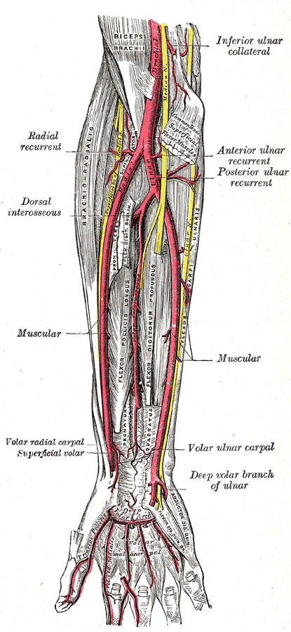
ULNAR ARTERY
SUMMARY
1. Arises from the bifurcation of the brachial artery in the cubital fossa.2. Exits the cubital fossa by passing deep to the pronator teres & FDS near the median nerve.
3. It then leaves the median nerve and lies on the FDP where it is joined by the ulnar nerve on its ulnar side.
4. The artery and nerve pass into the wrist on the flexor retinaculum (Guyon's canal), where the artery continues as the superficial palmar arch.
5. Surface marking: along a line medial to the biceps tendon in the cubital fossa to the pisiform.
BRANCHES
6. Anterior interosseous artery: lies on the interosseous membrane b/w the FDP & FPL. It supplies both muscles, interosseous membrane, radius & ulna. It then passes through the interosseous membrane at the level of the PQ.
7. Posterior interosseous artery: passes into the extensor compartment above the interosseous membrane.

Image: Gray, Henry. Anatomy of the Human Body. Philadelphia: Lea & Febiger, 1918; Bartleby.com, 2000. www.bartleby.com/107/ [Accessed 17 Apr. 2019].
Reference(s)
R.M.H McMinn (1998). Last’s anatomy: regional and applied. Edinburgh: Churchill Livingstone.
Gray, H., Carter, H.V. and Davidson, G. (2017). Gray’s anatomy. London: Arcturus.