Uncategorized files
From NeuroRehab.wiki
Showing below up to 50 results in range #1 to #50.
View (previous 50 | next 50) (20 | 50 | 100 | 250 | 500)
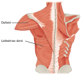
- 1119 Muscles that Move the Humerus b.png 1,038 × 960; 668 KB
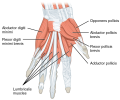
- 1121 Intrinsic Muscles of the Hand Deep LD.png 1,174 × 753; 370 KB
- 1121 Intrinsic Muscles of the Hand Superficial sin.png 1,224 × 1,023; 595 KB
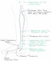
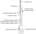
- 1123 Muscles of the Leg that Move the Foot and Toes c.png 648 × 1,238; 343 KB
- 1200px-Lungvolumes Updated.png 1,200 × 744; 84 KB
- 1920px-Standard deviation diagram.svg.png 1,920 × 960; 66 KB
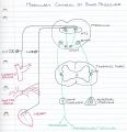
- 2019-08-16 190741.jpg 1,502 × 728; 205 KB
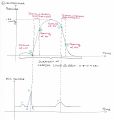
- 2019-08-16 191105.jpg 1,402 × 602; 148 KB
- 2019-08-20 055454.jpg 1,894 × 1,124; 338 KB
- 2019-09-04 194606.jpg 1,558 × 1,611; 352 KB
- 2019-09-12 062106.jpg 1,622 × 1,710; 199 KB
- 2019-09-24 192952.jpg 1,439 × 1,697; 231 KB
- 2022-11-16 220555.jpg 1,968 × 944; 267 KB
- 3-s2.0-B9780123851574007016-f00701-01-9780123851574.jpg 342 × 316; 21 KB
- 3-s2.0-B9781416055952000171-f17-01-9781416055952.jpg 508 × 347; 41 KB
- 37C2F5AB-0F2F-41DB-BE6A-413FF78EC83D.jpg 1,024 × 755; 125 KB
- 400px-Anterior Hip Muscles 2.PNG 400 × 600; 138 KB
- 460px-Leitner system alternative.svg.png 460 × 250; 15 KB
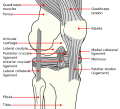
- 658px-Knee diagram.svg.png 658 × 600; 118 KB
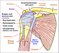
- 800px-Shoulder joint.svg.png 800 × 722; 259 KB
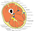
- 800px-Thigh cross section.svg.png 800 × 715; 214 KB
- 8544.jpg 225 × 375; 14 KB
- A04CB20C-6C01-4204-A812-80BD2D8FF0BC.jpg 1,024 × 704; 138 KB
- ACLS-Figure-30.jpg 1,686 × 560; 596 KB
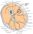
- Adductor canal.png 683 × 700; 238 KB
- Afo.jpg 380 × 513; 11 KB
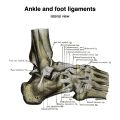
- Ankle-and-foot-ligaments-grays-illustrations.jpeg 1,600 × 1,600; 1.17 MB
- Anterior Hip Muscles 2.PNG 408 × 612; 98 KB
- Anterior tibial artery.png 200 × 600; 74 KB
- Atlas-grays-illustration-e343c75ad1817d356a16b18b6175b89b03d9b0ca.jpeg 1,600 × 1,117; 190 KB
- Atypical-thoracic-vertebrae-grays-illustration.jpeg 1,600 × 1,600; 717 KB
- Axillary-artery-diagrams.jpg 1,024 × 1,024; 266 KB
- Axis-grays-illustration-2.jpeg 1,240 × 1,359; 211 KB
- Axis-grays-illustration-3.jpeg 1,600 × 1,188; 188 KB
- B9781455710782000067 f006-015-9781455710782.jpg 560 × 342; 77 KB
- Biceps femoris muscle long head.PNG 368 × 1,000; 192 KB
- Blausen 0609 LegVeins.png 1,000 × 1,500; 781 KB
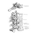
- Bones-and-ligaments-of-the-vertebral-column-illustrations-2.jpg 495 × 495; 113 KB
- Bones-and-ligaments-of-the-vertebral-column-illustrations-3.jpg 620 × 620; 140 KB
- Bones-and-ligaments-of-the-vertebral-column-illustrations.jpg 666 × 666; 96 KB
- Bony-pelvis-illustrations-2.jpg 614 × 614; 166 KB
- Bony-pelvis-illustrations.jpg 614 × 614; 163 KB
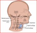
- C-spine surface anatomy.jpg 416 × 378; 30 KB
- Cavernous Sinus 2.png 500 × 377; 31 KB
- Cervical-ribs.JPEG 1,024 × 1,024; 363 KB
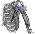
- Coracobrachialis.png 550 × 533; 233 KB
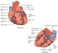
- Coronary-arteries-creative-commons-illustration.jpeg 2,283 × 2,096; 1.37 MB
- Costotransverse-joints-grays-illustration-2bca0209054a54d91549e3aac6dbd3e31f7d67c1.jpeg 1,600 × 1,138; 250 KB
- Costotransverse-joints-grays-illustration.jpeg 1,600 × 1,600; 868 KB
