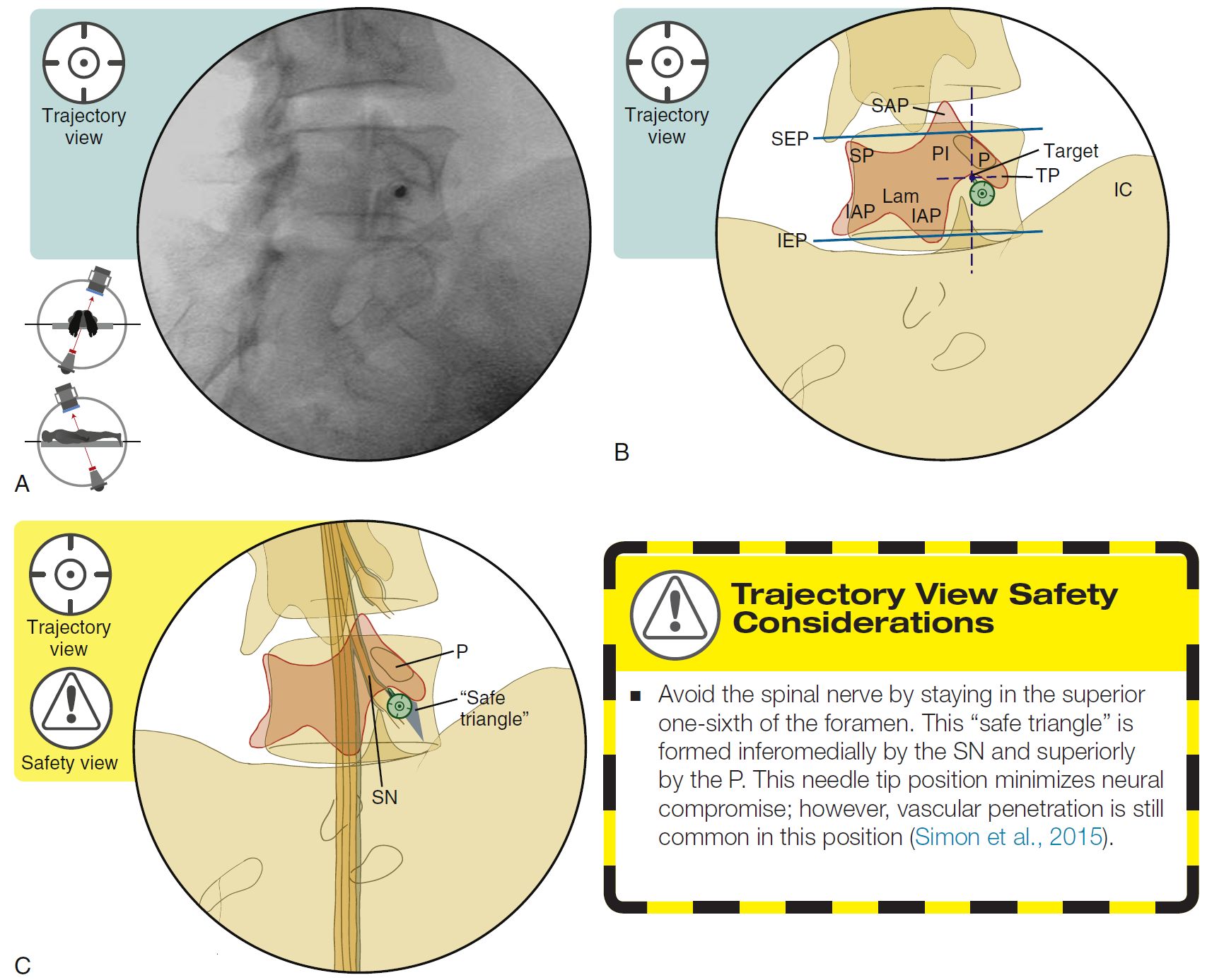
SPINE INJECTION-TRANSFORAMINAL BLOCK, TECHNIQUE
SUMMARY
1. Confirm the level.
2. Tilt the fluoroscope cephalad or caudad to line up the superior end plate (SEP) corresponding to the vertebra at which the injection is being performed.
3. Preferentially lining up the SEP rather than the inferior end plate (IEP) will favor an inferior-to-superior needle trajectory.
4. Oblique the fluoroscope ipsilateral to allow for proper visualization of the target point.
5. The target needle destination is just below the “chin” of the “Scotty dog” (i.e., adjacent to the pars interarticularis and inferior to the pedicle).
6. A more medial final needle tip placement requires a more oblique approach.
7. Identify a direct path to reach the target needle position.
8. Place the needle parallel to the fluoroscopic beam.

Image: Furman, Michael B., and Leland Berkwits. Atlas of Image-Guided Spinal Procedures. Elsevier, Inc, 2017.
Reference(s)
Furman, Michael B., and Leland Berkwits. Atlas of Image-Guided Spinal Procedures. Elsevier, Inc, 2017.
Horowitz AL. MRI Physics for Physicians. Springer Science & Business Media. (1989) ISBN:1468403338.
Mangrum W, Christianson K, Duncan S et-al. Duke Review of MRI Principles. Mosby. (2012) ISBN:1455700843.