MUSCLES-LATERAL COMPARTMENT OF LEG
SUMMARY
SUMMARY
1. Both muscles arise from the peroneal surface of the fibula, LONGUS above BREVIS.
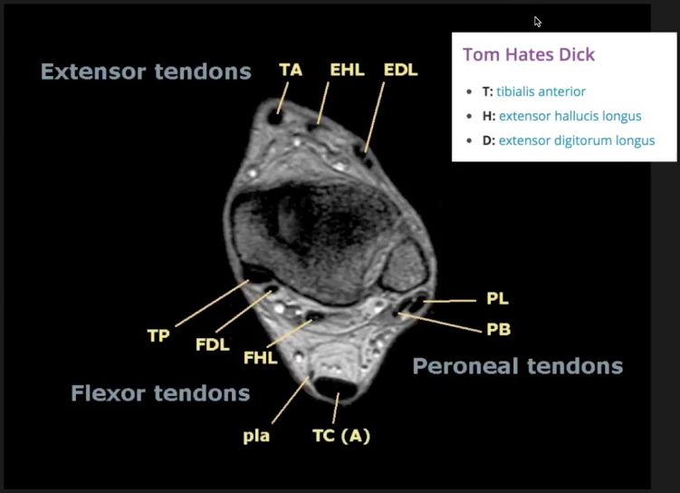
2. Both tendons groove the LATERAL malleolus, bound down by the superior peroneal retinaculum & at the peroneal trochlea of the calcaneus, bound down by the inferior peroneal retinaculum.
3. LONGUS crosses the sole of the foot to insert into the 1st MT base + medial cuniform.
4. BREVIS inserts into the tubercle at the 5th MT base.
5. Nerve supply - both muscles are supplied by the superficial peroneal nerve.
6. Arterial supply - perforating arteries of the ATA + peroneal a.
7. Actions - both muscles evert + plantar-flex the foot.
DETAILS
1. PERONEUS LONGUS - arises from the upper 2/3 of the peroneal surface of the fibula + head of the fibula + tib/fib joint + small area of lateral tibial condyle + intermuscular septa. The muscle lies behind the peroneus brevis and its tendon grooves behind the lateral malleolus. The tendon passes below the peroneal trochlea (of the calcaneus), obliquely across the sole of the foot to be inserted into the base of the 1st metatarsal + medial cuniform.
2. PERONEUS BREVIS - arises from the lower 1/3 of the fibula. The muscle lies in front of the peroneus longus and its tendon grooves behind the lateral malleolus. The tendon passes above the peroneal trochlea (of the calcaneus) to be inserted into the tubercle at the base of the 5th metatarsal bone.

Reference(s)
R.M.H McMinn (1998). Last’s anatomy: regional and applied. Edinburgh: Churchill Livingstone.
Gray, H., Carter, H.V. and Davidson, G. (2017). Gray’s anatomy. London: Arcturus.