MUSCLES-DEEP POSTERIOR COMPARTMENT OF LEG
SUMMARY
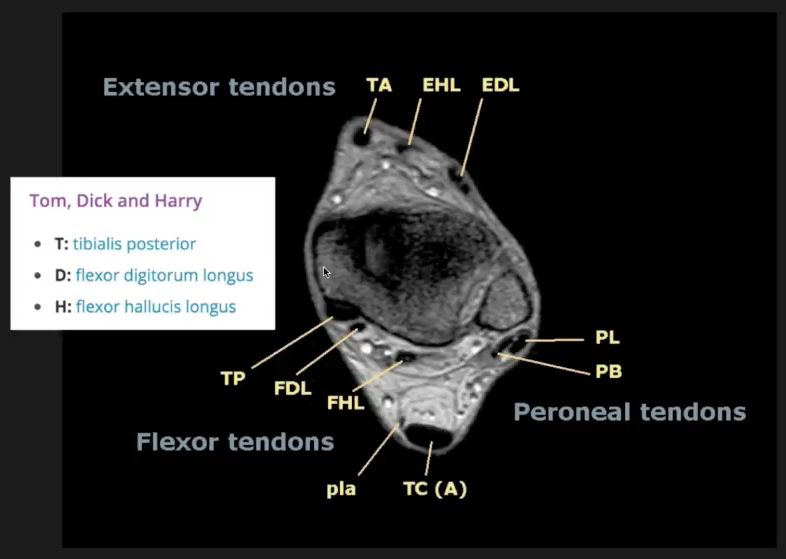
ALL TENDONS (EXCEPT THE POPLITEUS) PASS BENEATH THE MEDIAL FLEXOR RETINACULUM
1. TIBIALIS POSTERIOR - arises from the interosseous membrane, adjoining surfaces of tib/fib & adjoining aponeurosis of the FDL. The tendon grooves the back of the medial malleolus, passes above the sustentaculum tali to be inserted into the navicular tuberosity & 3 cuneiforms.
2. FLEXOR DIGITORUM LONGUS - tibial origin: posterior surface, below the soleal line. Fibular origin: by a broad aponeurosis. The tendon passes beneath the flexor retinaculum to enter the sole, where it crosses the FHL tendon to divide into 4 tendons. They pass into the fibrous flexor sheaths of the lateral 4 toes, perforate the tendons of the FDB to be inserted into the bases of the distal phalanges.
3. FLEXOR HALLUCIS LONGUS - arises from the flexor surface of the fibula, interosseous membrane & adjoining aponeurosis of the FDL. The tendon passes underneath the flexor retinaculum, grooves the posterior process of the talus & sustentaculum tali to be inserted into the base of the distal phalanx of the great toe.
4. POPLITEUS - arises from the popliteal surface of the tibia and inserts into a facet/pit in the lateral surface of the lateral femoral condyle (the tendon lies within the capsule of the knee joint: arcuate popliteal ligament) & lateral meniscus. This laterally rotates the femur on the fixed tibia & retracts the lateral meniscus.
5. All 4 muscles are supplied by the tibial nerve. Aretrial supply - posterior tibial & peroneal arteries.

Reference(s)
R.M.H McMinn (1998). Last’s anatomy: regional and applied. Edinburgh: Churchill Livingstone.
Gray, H., Carter, H.V. and Davidson, G. (2017). Gray’s anatomy. London: Arcturus.