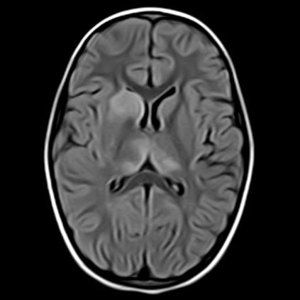
MRI-T1 OR T2 FLUID ATTENUATED SEQUENCE (FLAIR)
SUMMARY
1. Fluid Attenuated Sequence (FLAIR): to detect parenchymal oedema without the glaring high signal from CSF we suppress CSF.
2. At first glance FLAIR images appear similar to T1 (CSF is dark), the best way to tell the two apart is to look at the grey-white matter.
3. T1 sequences will have grey matter being darker than white matter.
4. T2 weighted sequences, whether fluid attenuated or not, will have white matter being darker than grey matter.

Image: The above is a T2w FLAIR, axial sequence through the brain demonstrating a hypointense lesion in the right caudate nucleus.
Reference(s)
Furman, Michael B., and Leland Berkwits. Atlas of Image-Guided Spinal Procedures. Elsevier, Inc, 2017.
Horowitz AL. MRI Physics for Physicians. Springer Science & Business Media. (1989) ISBN:1468403338.
Mangrum W, Christianson K, Duncan S et-al. Duke Review of MRI Principles. Mosby. (2012) ISBN:1455700843.