MRI-DIFFUSION WEIGHTED SEQUENCES (DWI)
SUMMARY
1. Diffusion weighted imaging assess the ease with which water molecules move around within a tissue (mostly representing fluid within the extracellular space) and gives insight into cellularity (e.g. tumours), cell swelling (e.g. ischaemia) and oedema.
2. Typically you will find three sets of images when diffusion weighted imaging is performed: DWI, ADC and B=0 images.
3. The dominant signal intensities of different tissues are:
- Fluid (e.g. urine, CSF): no restriction to diffusion
- Soft tissues (muscle, solid organs, brain): intermediate diffusion
- Fat: little signal due to paucity of water

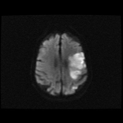
Image: The above is a DWI axial sequence through the brain demonstrating a (possible) ischemic infarct.
Reference(s)
Furman, Michael B., and Leland Berkwits. Atlas of Image-Guided Spinal Procedures. Elsevier, Inc, 2017.
Horowitz AL. MRI Physics for Physicians. Springer Science & Business Media. (1989) ISBN:1468403338.
Mangrum W, Christianson K, Duncan S et-al. Duke Review of MRI Principles. Mosby. (2012) ISBN:1455700843.