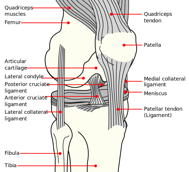
KNEE JOINT-TIBIAL OR MEDIAL COLLATERAL LIGAMENT
SUMMARY
1. Consists of superficial + deep parts.
2. Superficial part of the MCL - broad, flat band that extends from the medial femoral condyle to the subcutaneous surface of the proximal tibia.
3. It is separated from the tibial condyle by the semimembranosus tendon & bursa.
4. Below this it is separated from the proximal tibial shaft by the inferior genicular vessels & nerve.
5. The anterior margin of the superficial part lies free, whereas the posterior margin is attached to the medial meniscus.
6. The deep part of the MCL is a thickening of the capsule that attaches to the tibia and femur just beyond the articular margins & medial meniscus.

Image: Mysid [Public domain], via Wikimedia Commons [Accessed 13 Apr. 2019].
Reference(s)
R.M.H McMinn (1998). Last’s anatomy: regional and applied. Edinburgh: Churchill Livingstone.
Gray, H., Carter, H.V. and Davidson, G. (2017). Gray’s anatomy. London: Arcturus.