ILIOTIBIAL BAND
From NeuroRehab.wiki
SUMMARY
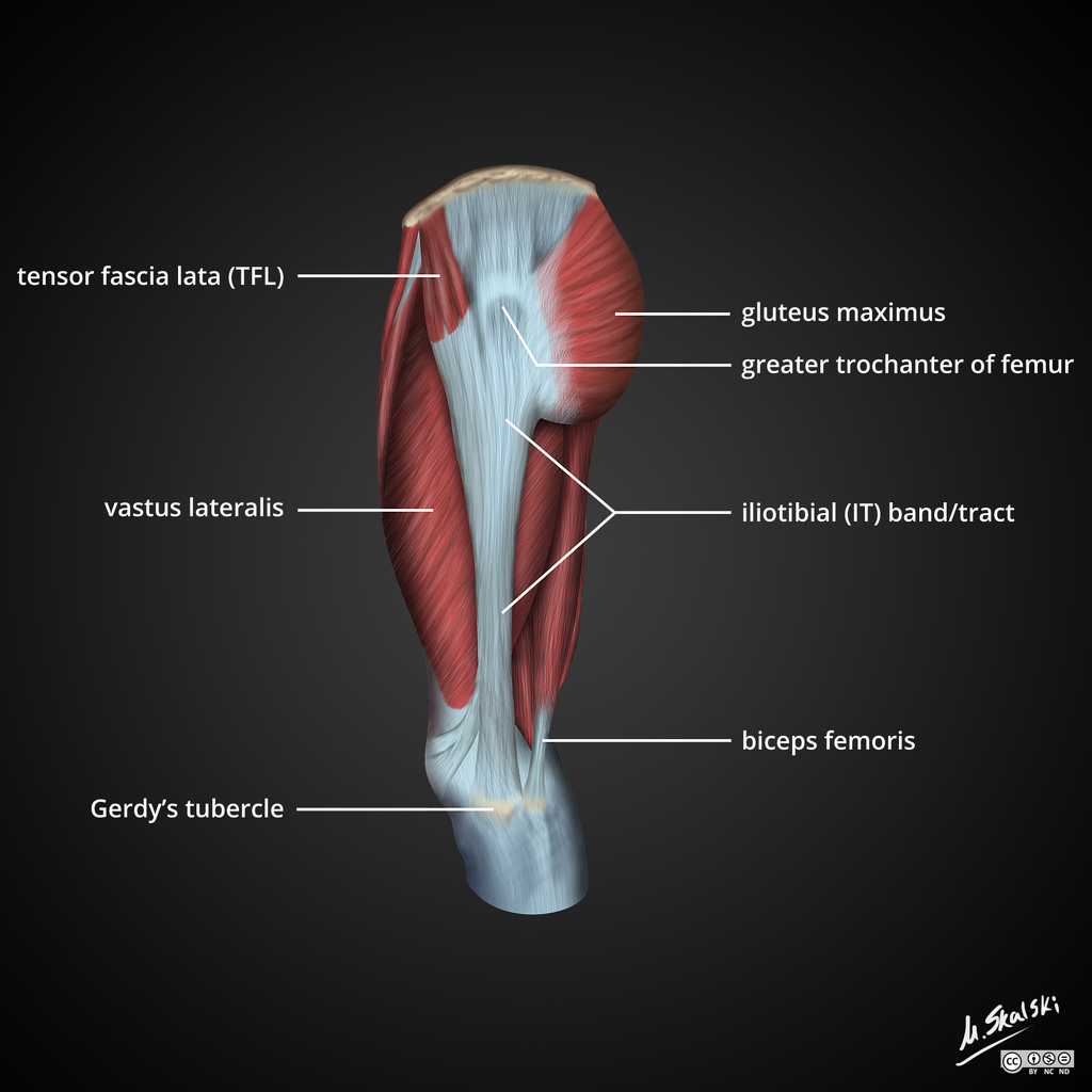
1. Thickening of the fascia lata that commences at the greater trochanter (where the gluteus maximus & tensor fasciae latae are inserted into it).
2. It passes vertically down the lateral aspect of the thigh, crosses the lateral condyle of the femur to be inserted into the anterior surface of the lateral tibial condyle (Gerdy's tubercle).
3. This maintains the knee in a hyperextended position + resists adduction at the hip.

Image: Case courtesy of Dr Matt Skalski, Radiopaedia.org. From the case rID: 36990
Reference(s)
R.M.H McMinn (1998). Last’s anatomy: regional and applied. Edinburgh: Churchill Livingstone.
Gray, H., Carter, H.V. and Davidson, G. (2017). Gray’s anatomy. London: Arcturus.