GOUT-RADIOGRAPHY
SUMMARY
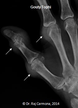
1. Tophi: typically located in the peri-articular area along the extensor surface, but maybe intra-articular or not associated with the joint at all.

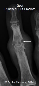
2. Punched out erosions: borders can be sclerotic because of the indolence of the process, creating a punched-out or "rat bitten" appearance.

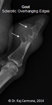
3. Sclerotic, overhanging edges: as the erosion develops, the edges of the cortex can be remodelled in an outward fashion, creating an overhanging edge.

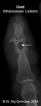
4. Intraosseous lesions: intraosseous tophi.

5. There is preserved joint space and no periarticular osteopenia.
Images: Carmona, R., 2014. Gout: Overview of X-Ray Findings. [online] Available at: RheumTutor. [Accessed 7 January 2022].
Reference(s)
Wilkinson, I., Furmedge, D. and Sinharay, R. (2017). Oxford handbook of clinical medicine. Oxford: Oxford University Press. Get it on Amazon.
Feather, A., Randall, D. and Waterhouse, M. (2020). Kumar And Clark’s Clinical Medicine. 10th ed. S.L.: Elsevier Health Sciences. Get it on Amazon.
Hannaman, R. A., Bullock, L., Hatchell, C. A., & Yoffe, M. (2016). Internal medicine review core curriculum, 2017-2018. CO Springs, CO: MedStudy.
Therapeutic Guidelines. Melbourne: Therapeutic Guidelines Limited. https://www.tg.org.au [Accessed 2021].