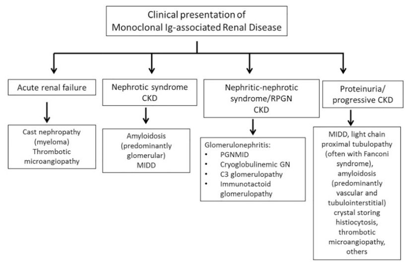
GLOMERULONEPHRITIS-MONOCLONAL Ig GN
SUMMARY
1. Characterised by monoclonal Ig deposits in the glomeruli and/or along the tubular basement membrane on immunofluorescence.
2. Ig can cause injury via: deposition, complement activation or precipitation/crystallization.
3. Diagnostic features on IF/IHC: proliferative forms of monoclonal Ig deposition disease, immunotactoid GN, fibrillary GN with monoclonal Ig deposits.
4. In the absence of these distinct patterns, GN with monotypic Ig glomerular deposits on IF/IHC and mesangial/capillary wall deposits on EM is labelled as proliferative GN with monoclonal Ig deposits.

RELATED DISEASES
6. MGUS: presence of an monoclonal Ig in the absence of plasma cell or lymphoid malignancy or end organ damage. Implies a benign condition. Defn: <10% bone marrow plasma cells, <3g/dL M protein, no evidence of myeloma.
7. MGRS: clonal plasma cell or B lymphocyte proliferation causing a renal lesion in the absence of haematologic malignancy. Defn: <10% bone marrow plasma cells, <3g/dL M protein, presence of renal lesions but absence of myeloma. Tx: directed at the clonal proliferation.
Image: Seethi et al (2018) The Complexity and Heterogeneity of Monoclonal Immunoglobulin-Associated Renal Diseases. JASN
Reference(s)
Wilkinson, I., Furmedge, D. and Sinharay, R. (2017). Oxford handbook of clinical medicine. Oxford: Oxford University Press. Get it on Amazon.
Feather, A., Randall, D. and Waterhouse, M. (2020). Kumar And Clark’s Clinical Medicine. 10th ed. S.L.: Elsevier Health Sciences. Get it on Amazon.
Hannaman, R. A., Bullock, L., Hatchell, C. A., & Yoffe, M. (2016). Internal medicine review core curriculum, 2017-2018. CO Springs, CO: MedStudy.
Therapeutic Guidelines. Melbourne: Therapeutic Guidelines Limited. https://www.tg.org.au [Accessed 2021].