CPPD DEPOSITION DISEASE-INVESTIGATION
SUMMARY
1. Diagnose CPPD by finding intracellular crystals that are blunted, rhomboid, and are weakly positively birefringent.
2. Joint-space narrowing.
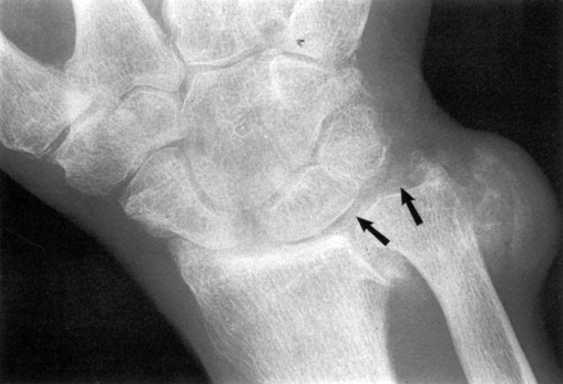
3. Chondrocalcinosis: ossification of the cartilage (see image below). Typical of pseudogout.

4. Cyst formation under the cartilage.
Reference(s)
Wilkinson, I., Furmedge, D. and Sinharay, R. (2017). Oxford handbook of clinical medicine. Oxford: Oxford University Press. Get it on Amazon.
Feather, A., Randall, D. and Waterhouse, M. (2020). Kumar And Clark’s Clinical Medicine. 10th ed. S.L.: Elsevier Health Sciences. Get it on Amazon.
Hannaman, R. A., Bullock, L., Hatchell, C. A., & Yoffe, M. (2016). Internal medicine review core curriculum, 2017-2018. CO Springs, CO: MedStudy.
Therapeutic Guidelines. Melbourne: Therapeutic Guidelines Limited. https://www.tg.org.au [Accessed 2021].