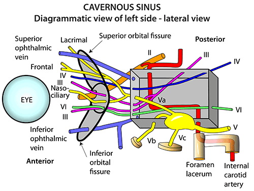
CN III (OCCULOMOTOR)-C. COURSE
SUMMARY
1. The nerve exits the medial surface at the base of the cerebral peduncle near the midline, just above the pons.
2. It passes between the PCA and SCA, then below & lateral to the PCOM, just below the free margin of the tentorium cerebelli.
3. It traverses the interpeduncular cistern below the floor of the third ventricle.
4. Lying below the optic tract, it enters the cavernous sinus by piercing the dura and arachnoid on its roof.
5. As it crosses the ICA it picks up sympathetic fibers from the ICA plexus (which originate from the T1 lateral horn cells) → for supply of the levator palpebrae superioris.
6. At the anterior pole of the cavernous sinus it divides into: superior & inferior divisions, which pass through the Annulus of Zihn to innervate the extraocular muscles.

Image: Whitaker, R. and Borley, N. (2016). Instant anatomy. 6th ed. Chichester (West Sussex): Wiley Blackwell, p.93.
Reference(s)
R.M.H McMinn (1998). Last’s anatomy: regional and applied. Edinburgh: Churchill Livingstone. Get it on Amazon.
Drake, Richard L., et al. Gray's Anatomy for Students. Elsevier, 2023. Get it on Amazon.