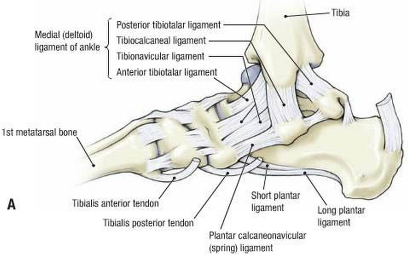
ANKLE JOINT-MEDIAL DELTOID LIGAMENT
SUMMARY
1. The deltoid ligament is a strong, flat, triangular band attached above to the apex, anterior & posterior borders of the medial malleolus.
SUPERFICIAL FIBERS
2. Tibionavicular pass forward to be inserted into the tuberosity of the navicular bone, and immediately behind this it blends with the medial margin of the plantar calcaneonavicular ligament (spring ligament).
3. Tibiocalcaneal descend almost perpendicularly to be inserted into the whole length of the sustentaculum tali of the calcaneus.
4. Posterior tibiotalar pass backward and laterally to be attached to the inner side of the talus, and to the prominent tubercle on its posterior surface, medial to the groove for the tendon of the flexor hallucis longus.
DEEP FIBERS
5. Anterior tibiotalar fibers are attached to the anterior colliculus of the medial malleolus, and below to the anteromedial talus.

Image: ANATOMY BODY DIAGRAM. (2019). ANATOMY BODY DIAGRAM - Anatomy of Reproductive, Muscle, Inner Body and etc.. [online] Available at: https://bloginonline.com/ [Accessed 13 Apr. 2019].
Reference(s)
R.M.H McMinn (1998). Last’s anatomy: regional and applied. Edinburgh: Churchill Livingstone.
Gray, H., Carter, H.V. and Davidson, G. (2017). Gray’s anatomy. London: Arcturus.