Difference between revisions of "KNEE JOINT-TIBIAL OR MEDIAL COLLATERAL LIGAMENT"
(Imported from text file) |
(Imported from text file) |
||
| (One intermediate revision by the same user not shown) | |||
| Line 1: | Line 1: | ||
[[Summary Article| | ===== [[Summary Article|'''SUMMARY''']] ===== | ||
1. Consists of <i>superficial + deep parts.</i> | |||
<br/>2. S<i>uperficial part of the MCL - | <br/>2. S<i>uperficial part of the MCL - </i>broad, flat band that extends from the medial femoral condyle to the subcutaneous surface of the proximal tibia. | ||
<br/>3. It is separated from the tibial condyle by the semimembranosus tendon & bursa. | <br/>3. It is separated from the tibial condyle by the semimembranosus tendon & bursa. | ||
<br/>4. Below this it is separated from the proximal tibial shaft by the inferior genicular vessels & nerve. | <br/>4. Below this it is separated from the proximal tibial shaft by the inferior genicular vessels & nerve. | ||
<br/>5. The <i>anterior margin </i>of the superficial part lies free, whereas the <i>posterior margin </i>is attached to the medial meniscus. | <br/>5. The <i>anterior margin </i>of the superficial part lies free, whereas the <i>posterior margin </i>is attached to the medial meniscus. | ||
<br/>6. The <i>deep part of the MCL </i>is a thickening of the capsule | <br/>6. The <i>deep part of the MCL </i>is a thickening of the capsule that attaches to the tibia and femur just beyond the articular margins & medial meniscus. | ||
<br/> | |||
<br/>[[Image:658px-Knee_diagram.svg.png]] | <br/>[[Image:658px-Knee_diagram.svg.png]] | ||
<br/> | <br/> | ||
<br/><b>Image:</b> | <br/><b>Image:</b> Mysid [Public domain], [https://commons.wikimedia.org/wiki/File:Knee_diagram.svg via Wikimedia Commons] [Accessed 13 Apr. 2019]. | ||
==Reference(s)== | ==Reference(s)== | ||
Latest revision as of 11:29, 1 January 2023
SUMMARY
1. Consists of superficial + deep parts.
2. Superficial part of the MCL - broad, flat band that extends from the medial femoral condyle to the subcutaneous surface of the proximal tibia.
3. It is separated from the tibial condyle by the semimembranosus tendon & bursa.
4. Below this it is separated from the proximal tibial shaft by the inferior genicular vessels & nerve.
5. The anterior margin of the superficial part lies free, whereas the posterior margin is attached to the medial meniscus.
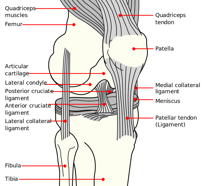
6. The deep part of the MCL is a thickening of the capsule that attaches to the tibia and femur just beyond the articular margins & medial meniscus.

Image: Mysid [Public domain], via Wikimedia Commons [Accessed 13 Apr. 2019].
Reference(s)
R.M.H McMinn (1998). Last’s anatomy: regional and applied. Edinburgh: Churchill Livingstone.
Gray, H., Carter, H.V. and Davidson, G. (2017). Gray’s anatomy. London: Arcturus.