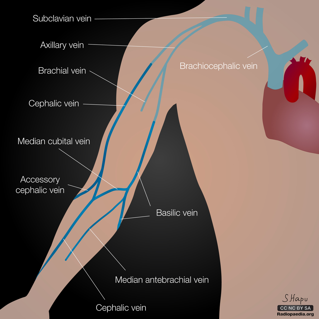
AXILLARY VEIN
SUMMARY
1. Origin - formed by the union of the venae comitantes of the brachial a. & basilic v.
2. Location - courses medial to the axillary a. in the axilla. Leaves the axilla by passing through the apex, anterior to the subclavian a.
3. Divisions - divided into 3 parts by the PECTORALIS MINOR. First part (above), second part (behind), third part (below).
4. Drainage - upper limb, axilla, superolateral chest wall.
5. Termination - over the upper surface of first rib, anterior to SCALENUS ANTERIOR it becomes the subclavian v.

Image: Case courtesy of Dr Sachintha Hapugoda, Radiopaedia.org. From the case rID: 52482
Reference(s)
R.M.H McMinn (1998). Last’s anatomy: regional and applied. Edinburgh: Churchill Livingstone.
Gray, H., Carter, H.V. and Davidson, G. (2017). Gray’s anatomy. London: Arcturus.