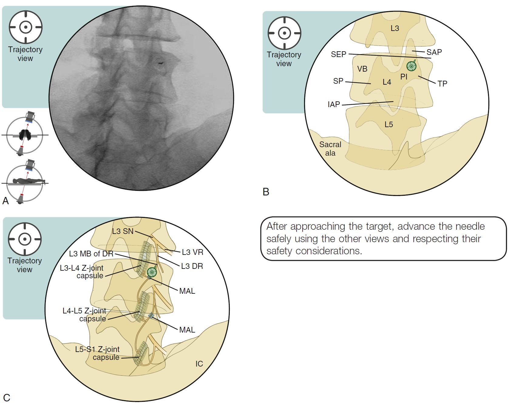
SPINE INJECTION-FACET JOINT RFA, TECHNIQUE
SUMMARY
1. Confirm the level.
2. Oblique and tilt the C-arm image intensifier to obtain an optimal AP view with the spinous process (SP) at midline and squaring off the superior end plate (SEP) of the vertebral body.
3. Oblique the C-arm about 20o toward the symptomatic side (do not oblique for the L5 dorsal ramus).
4. Tilt the C-arm about 40-45o caudally from the squared SEP.
5. This angle is used for entry and to approximate the trajectory of the target nerve for a “parallel placement” along the nerve.
6. The target is the lateral border of the SAP and the small concavity that is formed by the junction of SAP and the TP.
7. At the L5 level, there is only a rudimentary MAL, and the iliac crest will interfere with oblique needle positioning. Therefore, the oblique angle will be close to 0o.
8. As this is the trajectory view, the needle entry position should be parallel to the C-arm beam.

Image: Furman, Michael B., and Leland Berkwits. Atlas of Image-Guided Spinal Procedures. Elsevier, Inc, 2017.
Reference(s)
Furman, Michael B., and Leland Berkwits. Atlas of Image-Guided Spinal Procedures. Elsevier, Inc, 2017.
Horowitz AL. MRI Physics for Physicians. Springer Science & Business Media. (1989) ISBN:1468403338.
Mangrum W, Christianson K, Duncan S et-al. Duke Review of MRI Principles. Mosby. (2012) ISBN:1455700843.