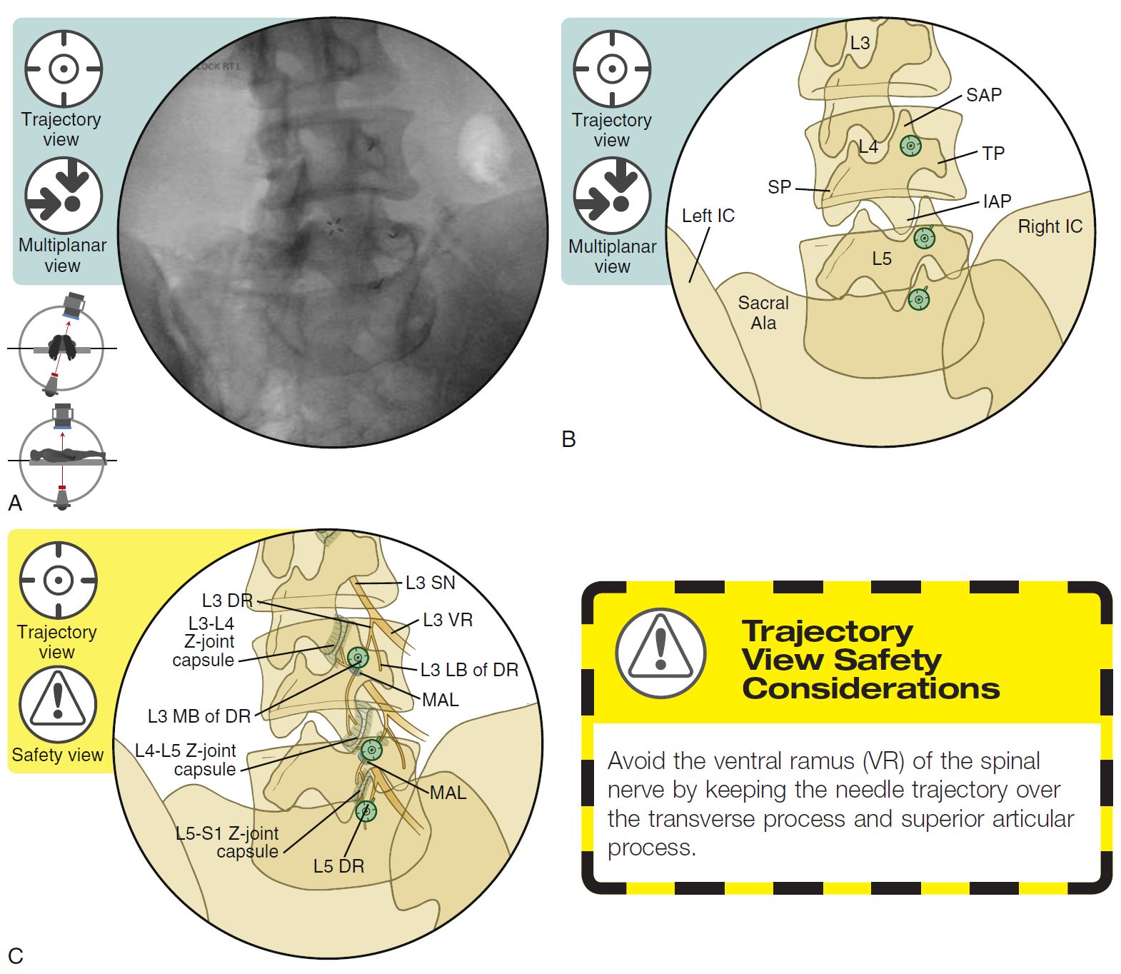
SPINE INJECTION-FACET JOINT MBB, TECHNIQUE
SUMMARY
1. Confirm the level with an AP view.
2. Tilt the fluoroscope’s image intensifier to line up the superior end plate of the targeted segment.
3. Oblique the C-arm image intensifier ipsilaterally to form the “Scotty dog” and optimize visualization of the junction of the transverse process (TP) and superior articular process (SAP).
4. For the L1 to L4 medial branches: aim for the junction of the SAP and TP, where the target nerve crosses midway between the superior border of the TP and mamillo-accessory ligament (MAL) notch (“Eye of the Scotty dog").
5. The L5 dorsal ramus: aim for the middle of the base of the SAP, slightly below the sacral ala. If the iliac crest interferes with needle placement, oblique the fluoroscope 5o to 10o back toward AP to visualize a non-obstructed trajectory to the junction of the SAP and sacral ala.
6. Both of the nerves that innervate each targeted lumbar facet joint will need to be anesthetized.
7. The needle should be placed parallel to the fluoroscopic beam in this trajectory view.

Image: Furman, Michael B., and Leland Berkwits. Atlas of Image-Guided Spinal Procedures. Elsevier, Inc, 2017.
Reference(s)
Furman, Michael B., and Leland Berkwits. Atlas of Image-Guided Spinal Procedures. Elsevier, Inc, 2017.
Horowitz AL. MRI Physics for Physicians. Springer Science & Business Media. (1989) ISBN:1468403338.
Mangrum W, Christianson K, Duncan S et-al. Duke Review of MRI Principles. Mosby. (2012) ISBN:1455700843.