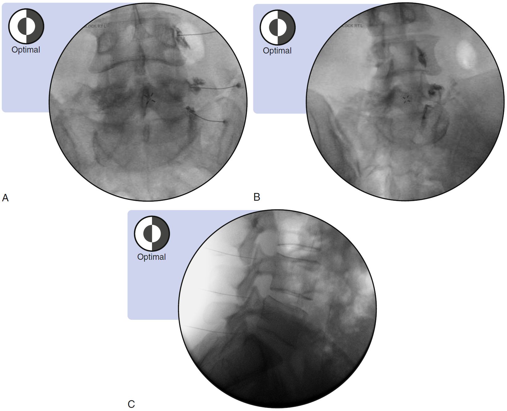
SPINE INJECTION-FACET JOINT MBB, OPTIMAL VIEWS
SUMMARY
1. L1 to L4 medial branches: Contrast flow will smoothly outline the medial border, thereby indicating the spread of contrast along the lateral surface of the base of the superior articular process (SAP), without epidural or vascular flow.
2. L5 dorsal ramus: Contrast flow will form a smooth margin around the base of the SAP of the sacrum, without epidural or vascular flow.

Image: Furman, Michael B., and Leland Berkwits. Atlas of Image-Guided Spinal Procedures. Elsevier, Inc, 2017.
Reference(s)
Furman, Michael B., and Leland Berkwits. Atlas of Image-Guided Spinal Procedures. Elsevier, Inc, 2017.
Horowitz AL. MRI Physics for Physicians. Springer Science & Business Media. (1989) ISBN:1468403338.
Mangrum W, Christianson K, Duncan S et-al. Duke Review of MRI Principles. Mosby. (2012) ISBN:1455700843.