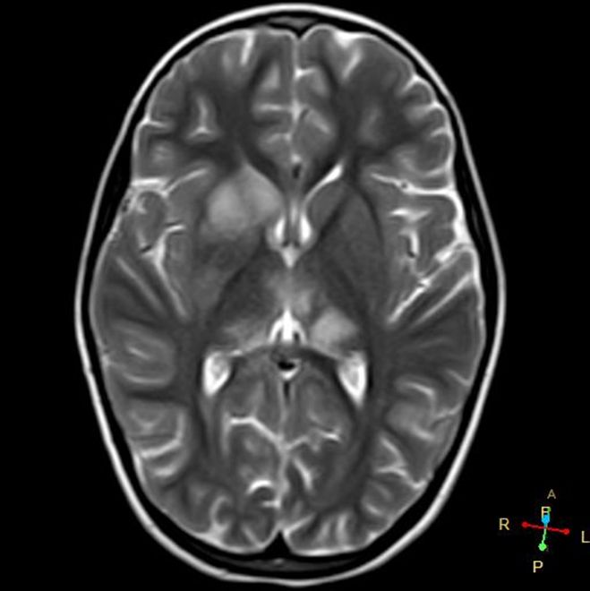
MRI-T2 SEQUENCE
SUMMARY
T2 weighted (T2W) sequences are part of almost all MRI protocols. Without modification the dominant signal intensities of different tissues are:
- Fluid (e.g. urine, CSF): high signal intensity (white)
- Muscle: intermediate signal intensity (grey)
- Fat: high signal intensity (white)
- Brain, grey matter: intermediate signal intensity (grey)
- Brain, white matter: hypointense compared to grey matter (dark)

Image: The above is a T2W axial sequence of the brain.
Reference(s)
Furman, Michael B., and Leland Berkwits. Atlas of Image-Guided Spinal Procedures. Elsevier, Inc, 2017.
Horowitz AL. MRI Physics for Physicians. Springer Science & Business Media. (1989) ISBN:1468403338.
Mangrum W, Christianson K, Duncan S et-al. Duke Review of MRI Principles. Mosby. (2012) ISBN:1455700843.