MRI-T1 SEQUENCE
SUMMARY
1. T1 weighted (T1W) sequences are part of almost all MRI protocols and are best thought of as the most 'anatomical' of images.
2. Historically the T1W sequence was known as the anatomical sequence, resulting in images that most closely approximate the appearances of tissues macroscopically, although even this is a gross simplification.
3. The dominant signal intensities of different tissues are:
- Fluid (e.g. urine, CSF): low signal intensity (black)
- Muscle: intermediate signal intensity (grey)
- Fat: high signal intensity (white)
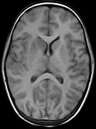
- Brain, grey matter: intermediate signal intensity (grey)
- Brain, white matter: hyperintense compared to grey matter (whitish)

Image: The above image is a T1W axial sequence of the brain.
Reference(s)
Furman, Michael B., and Leland Berkwits. Atlas of Image-Guided Spinal Procedures. Elsevier, Inc, 2017.
Horowitz AL. MRI Physics for Physicians. Springer Science & Business Media. (1989) ISBN:1468403338.
Mangrum W, Christianson K, Duncan S et-al. Duke Review of MRI Principles. Mosby. (2012) ISBN:1455700843.