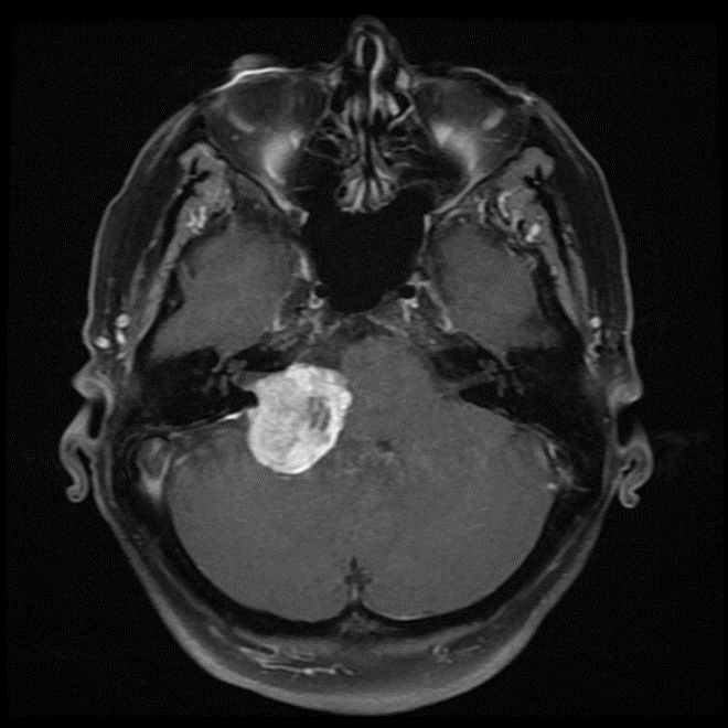
MRI-T1 CONTRAST SEQUENCE
SUMMARY
1. The most commonly used contrast agents in MRI are gadolinium based.
2. At the concentrations used, these agents have the effect of causing T1 signal to be increased (sometimes referred to as T1 shortening).
3. The contrast is injected intravenously (typically 5-15 mL) and scans are obtained a few minutes after administration.
4. Pathological tissues (tumours, areas of inflammation/infection) will demonstrate accumulation of contrast (mostly due to leaky blood vessels) and therefore appear as brighter than surrounding tissue.
5. Often post contrast T1 sequences are also fat suppressed to make this easier to appreciate.

Image: The above is a T1 contrast enhanced axial sequence of the brain demonstrating a contrast enhancing lesion.
Reference(s)
Furman, Michael B., and Leland Berkwits. Atlas of Image-Guided Spinal Procedures. Elsevier, Inc, 2017.
Horowitz AL. MRI Physics for Physicians. Springer Science & Business Media. (1989) ISBN:1468403338.
Mangrum W, Christianson K, Duncan S et-al. Duke Review of MRI Principles. Mosby. (2012) ISBN:1455700843.