MRI-SUSCEPTIBILITY WEIGHTED IMAGING (SWI)
From NeuroRehab.wiki
Revision as of 10:30, 24 July 2023 by Dr Appukutty Manickam (talk | contribs) (Imported from text file)
SUMMARY
1. Susceptibility weighted imaging (SWI) can be used to detect blood products and calcium.
2. Generally these sequences exploit what is referred to as T2* (T2 star) which is highly sensitive to small perturbations in the local magnetic field.
3. The most sensitive of these sequences is SWI and is also able to distinguish calcium from blood.

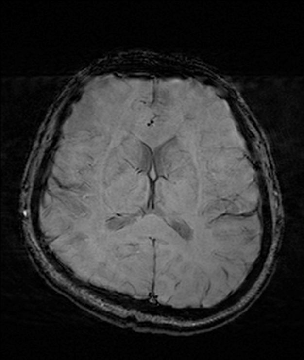
Image: The above is a SWI axial sequence through the brain.
Reference(s)
Furman, Michael B., and Leland Berkwits. Atlas of Image-Guided Spinal Procedures. Elsevier, Inc, 2017.
Horowitz AL. MRI Physics for Physicians. Springer Science & Business Media. (1989) ISBN:1468403338.
Mangrum W, Christianson K, Duncan S et-al. Duke Review of MRI Principles. Mosby. (2012) ISBN:1455700843.