MRI-MR SPECTROSCOPY
From NeuroRehab.wiki
Revision as of 10:30, 24 July 2023 by Dr Appukutty Manickam (talk | contribs) (Imported from text file)
SUMMARY
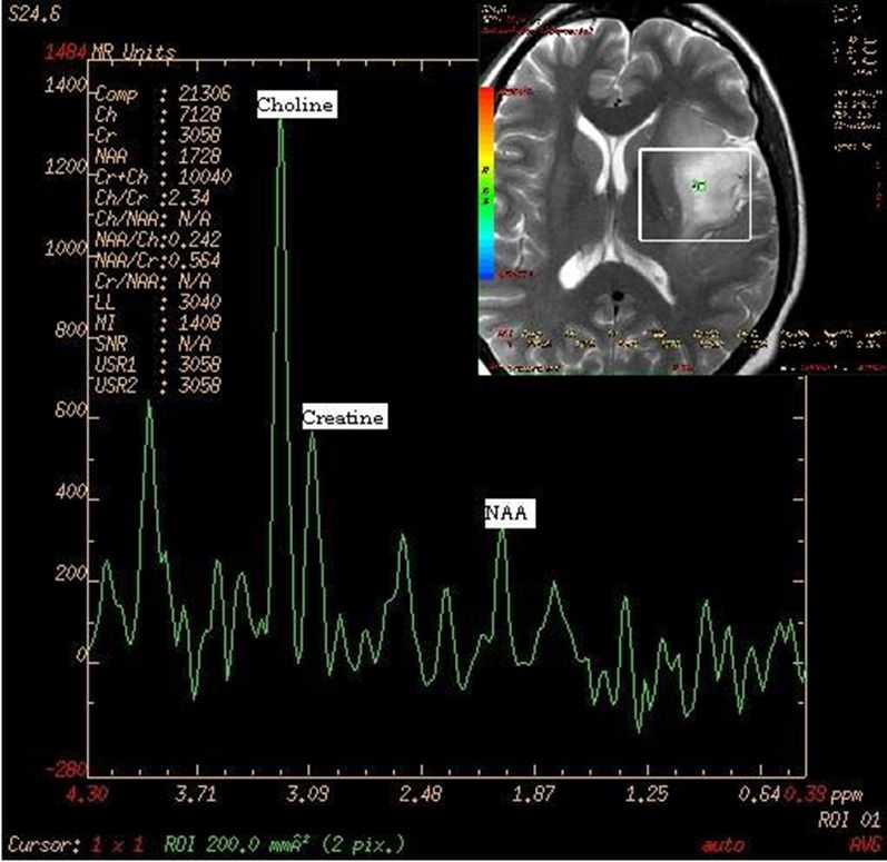
1. Different compounds interact with the magnetic field of MRI scanners slightly differently and the amounts of these compounds can be detected in a quantifiable way in a prescribed region of tissue.
2. These can be used to help characterise the tissue to aid in diagnosis or grading of tumours.

Image: MR spectrogram.
Reference(s)
Furman, Michael B., and Leland Berkwits. Atlas of Image-Guided Spinal Procedures. Elsevier, Inc, 2017.
Horowitz AL. MRI Physics for Physicians. Springer Science & Business Media. (1989) ISBN:1468403338.
Mangrum W, Christianson K, Duncan S et-al. Duke Review of MRI Principles. Mosby. (2012) ISBN:1455700843.