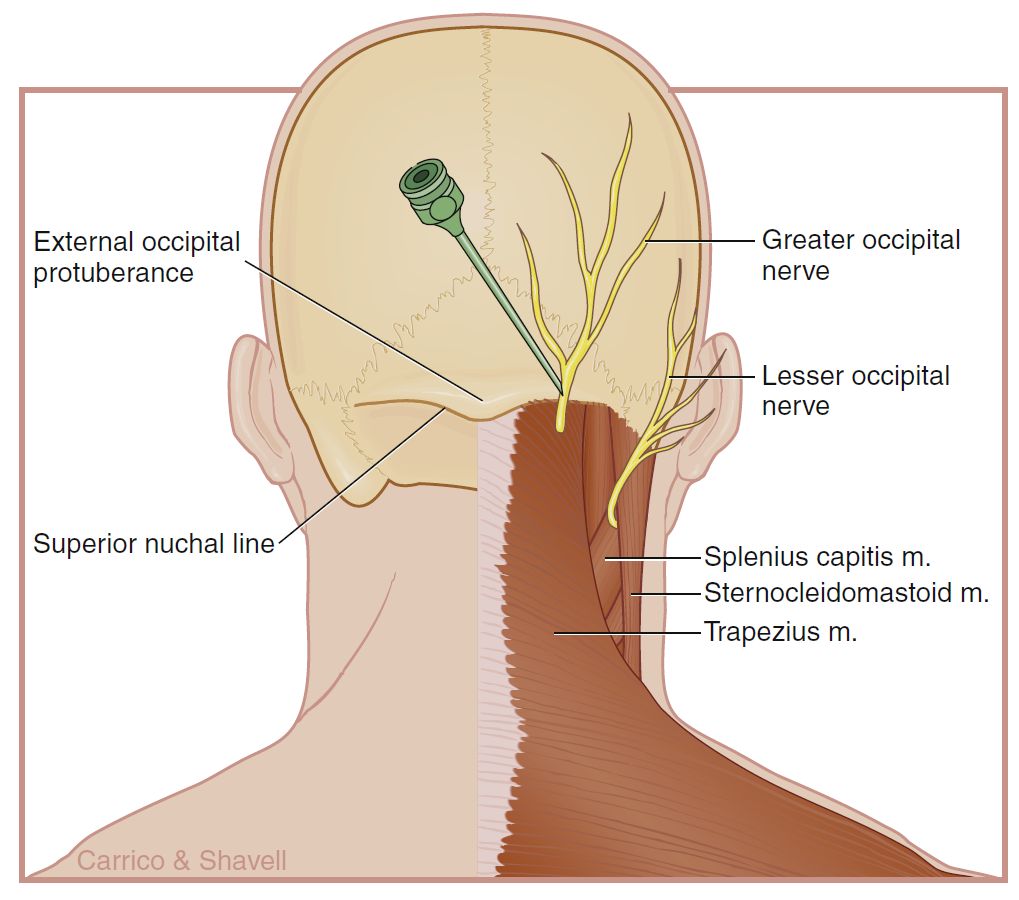
GREATER OCCIPITAL NERVE-INJECTION
SUMMARY
1. The patient is placed in a sitting position with the neck flexed and the forehead on a padded bedside table.
2. The occipital artery is then palpated at the level of the superior nuchal ridge.
3. First block: a total of 4 mL of local anesthetic is drawn up in a 12-mL sterile syringe with 80 mg of depot steroid; 40 mg of depot steroid is added with subsequent blocks.
4. The occipital artery is then palpated at the level of the superior nuchal ridge.
5. A high-frequency linear US transducer is placed in the transverse position at the nuchal ridge at the point at which the pulsation of the occipital artery was identified.
6. The occipital nerve will be in close proximity to the artery and will appear as a hypoechoic ovoid-shaped structure that does not compress when pressure is applied.
7. A 3½-inch spinal needle is inserted at the medial border of the US transducer using an in-plane approach and advanced toward the nerve until the tip impinges on the occipital periosteum.
8. When the needle tip is in proximity to the greater occipital nerve, after careful aspiration, 4 mL of the injectate is placed while injecting in a fan-like manner.
9. When the occipital nerve is injected, care must be taken to avoid entering the foramen magnum.

Image: Waldman, Steven D. Atlas of Pain Management Injection Techniques: E-Book. Elsevier Health Sciences, 13 July 2022.
Reference(s)
Furman, Michael B., and Leland Berkwits. Atlas of Image-Guided Spinal Procedures. Elsevier, Inc, 2017.
Horowitz AL. MRI Physics for Physicians. Springer Science & Business Media. (1989) ISBN:1468403338.
Mangrum W, Christianson K, Duncan S et-al. Duke Review of MRI Principles. Mosby. (2012) ISBN:1455700843.