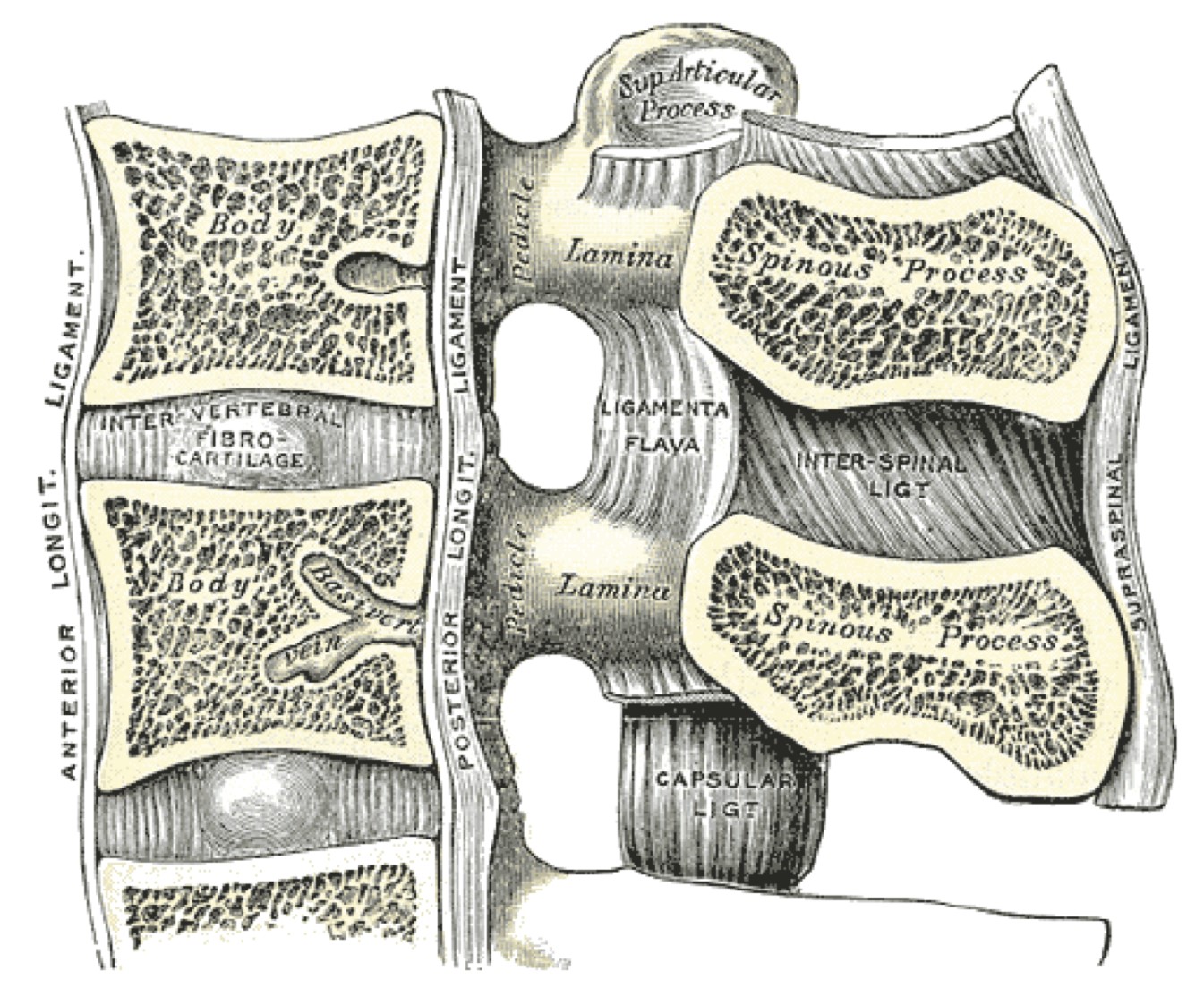
VERTEBRAL JOINTS-LONGITUDINAL LIGAMENTS
SUMMARY
1. Anterior longitudinal ligament: extends from the anterior tubercle of the atlas (‘front’ or ALL is attached to C1, ‘back’ or PLL is attached to C2) to the upper part of the sacrum
- It is attached firmly to the periosteum of the vertebral bodies but less so over the discs
2. Posterior longitudinal ligament: extends from the back of the body of the axis (‘front’ or ALL is attached to C1, ‘back’ or PLL is attached to C2) to the sacral canal
- It is firmly attached to the discs and loosely attached to the vertebral bodies
- This is to allow free exit of the basivertebral veins that exit the back of the vertebral bodies. At the top it is continued as the tectorial membrane above C1

Image: Case courtesy of Craig Hacking, Radiopaedia.org. From the case rID: 83584.
Reference(s)
R.M.H McMinn (1998). Last’s anatomy: regional and applied. Edinburgh: Churchill Livingstone. Get it on Amazon.
Drake, Richard L., et al. Gray's Anatomy for Students. Elsevier, 2023. Get it on Amazon.