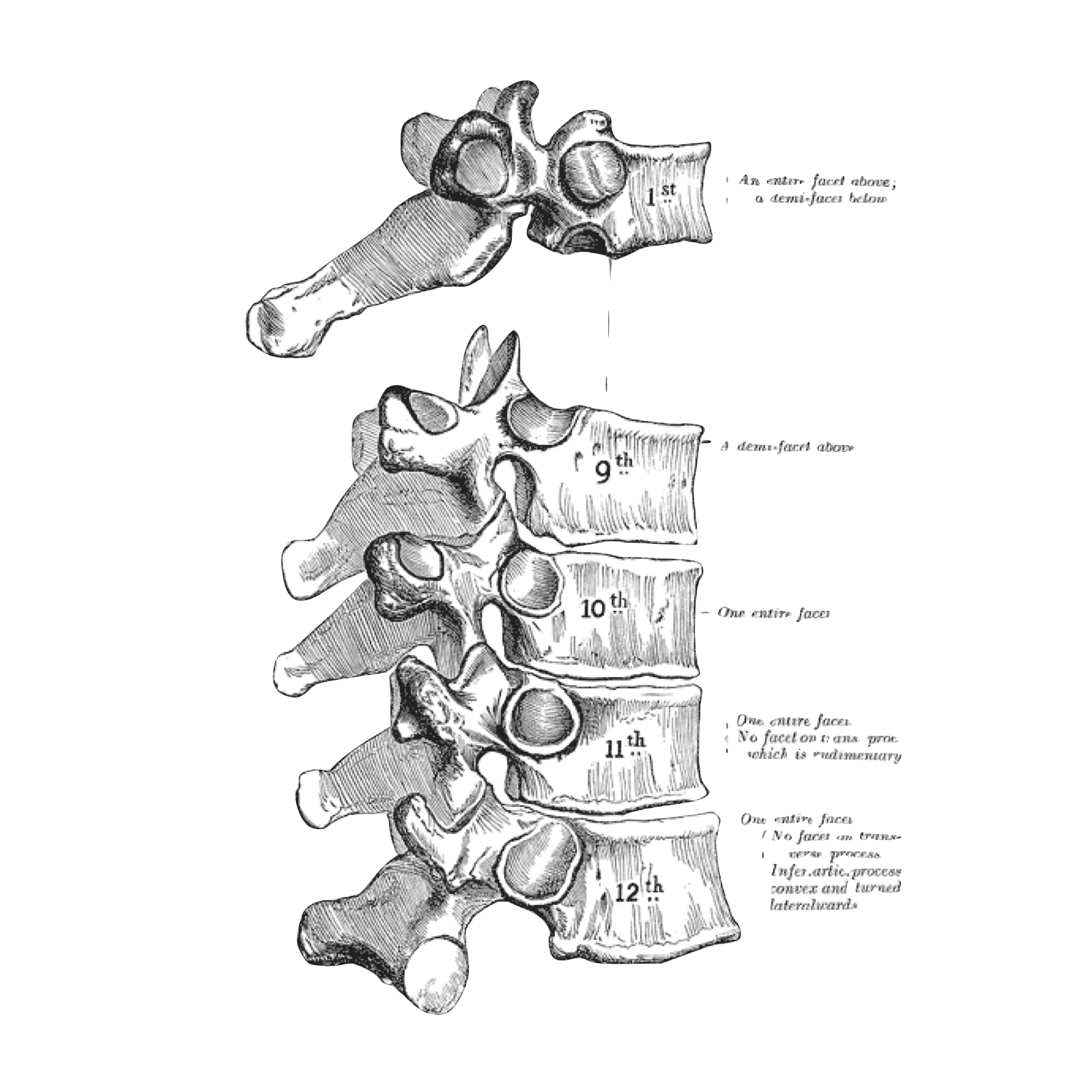
THORACIC VERTEBRA-T11 & T12
From NeuroRehab.wiki
Revision as of 12:35, 26 March 2023 by Dr Appukutty Manickam (talk | contribs) (Imported from text file)
SUMMARY
1. These carry single costal facets for their respective ribs
2. T11 costal facet lies behind the body & upper part of the pedicle
3. T12 costal facet lies near the lower margin
4. The TP is stunted and its base projects upwards into a rounded mamillary process & downwards into a sharp accessory tubercle

Image: Case courtesy of Craig Hacking, Radiopaedia.org. From the case rID: 82887
Reference(s)
R.M.H McMinn (1998). Last’s anatomy: regional and applied. Edinburgh: Churchill Livingstone. Get it on Amazon.
Drake, Richard L., et al. Gray's Anatomy for Students. Elsevier, 2023. Get it on Amazon.