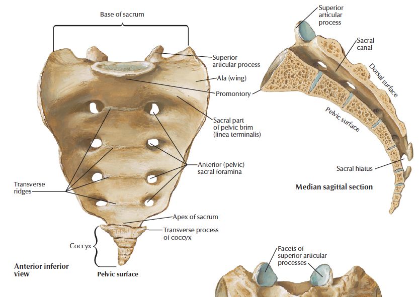
SACRUM-PELVIC SURFACE OSTEOLOGY
SUMMARY
1. Concave surface, smooth.
2. 4 transverse ridges persist to mark the lines of ossification. These represent the intervertebral discs.
3. On each side, the 4 anterior sacral foramina become smaller from above down, the rounded bars of bone between them represent fused head & neck of ribs.
4. The medial boundaries of the anterior sacral foramina are formed by the vertebral bodies.
5. The ala of the sacrum projects laterally from the upper surface of S1 vertebra, its margin forms the pelvic brim.
6. The mass of bone lateral to the foramina, the lateral mass, is formed by the fusion of the costal elements. It is indented by grooves for the anterior rami of S1-4.

Image: Sacrum. Netter. (2014). Atlas of Human Anatomy, Sixth Edition. 6th ed. Elsevier.
Reference(s)
R.M.H McMinn (1998). Last’s anatomy: regional and applied. Edinburgh: Churchill Livingstone. Get it on Amazon.
Drake, Richard L., et al. Gray's Anatomy for Students. Elsevier, 2023. Get it on Amazon.