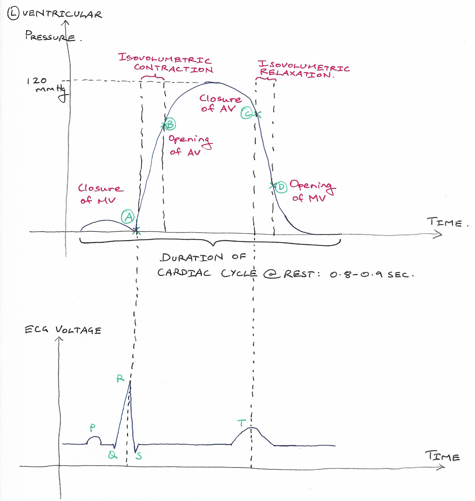
CARDIAC ELECTRICAL ACTIVITY-RELATIVE TO CYCLE
SUMMARY
1. P wave: atrial depolarization. Atrial systole begins after the P wave.
2. QRS complex: ventricular depolarization. Ventricular systole begins near the end of the R wave.
3. T wave: ventricular repolarization. Ventricular systole ends after the T wave.
4. Other waves: J wave (junction of S wave & ST segment, common in hypothermia), U wave (low voltage wave after T wave, common in hypokalemia).
NB: Since the T wave represents repolarization, one would expect the deflection to be negative. However, repolarization occurs from epicardium to endocardium, opposite of depolarization. Thus, the deflection is in the same direction as depolarization.

Image: Dr. Appukutty Manickam.
Reference(s)
Barrett, K.E., Barman, S.M., Brooks, H.L., X, J. and Ganong, W.F. (2019). Ganong’s review of medical physiology. 26th ed. New York: Mcgraw-Hill Education