SHOULDER JOINT
SUMMARY
GLENOHUMERAL JOINT
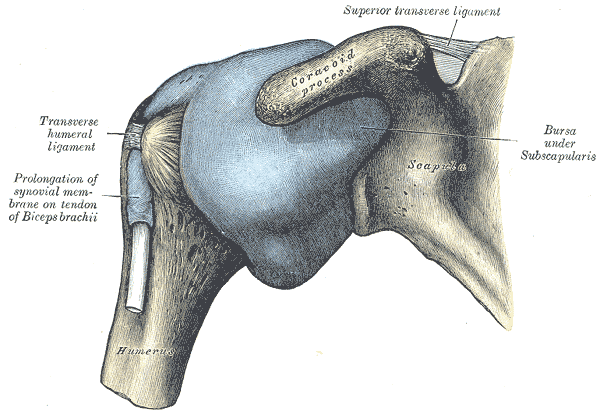
1. Synovial ball and socket joint, between the glenoid of the scapula & humeral head.
ARTICULAR SURFACES & CAPSULE
2. A ring of fibrocartilage, the glenoid labrum attached to the margins of the glenoid cavity, deepens the fossa.
3. The capsule of the joint is attached to the margins of the labrum & anatomical neck of the humerus (except inferiorly, where it is attached to the surgical neck).
LIGAMENTS & BURSAE
4. The joint is reinforced by the gleno-humeral ligaments (superior, middle, inferior), coraco-humeral ligament, coraco-acromial ligament.
5. The large subacromial bursa lies underneath the coraco-acromial ligament, over the supraspinatus tendon. This bursa never communicates with the shoulder joint, unless if the supraspinatus is torn.
6. Subacromial bursitis => tenderness over the greater tuberosity of the humerus, under the deltoid & disappears when the arm is abducted.
NERVE SUPPLY
7. Nerve supply: axillary n., musculocutaneous n. & suprascapular n.

Image: Gray, Henry. Anatomy of the Human Body. Philadelphia: Lea & Febiger, 1918; Bartleby.com, 2000. www.bartleby.com/107/ [Accessed 15 Nov. 2018].
Reference(s)
R.M.H McMinn (1998). Last’s anatomy: regional and applied. Edinburgh: Churchill Livingstone.
Gray, H., Carter, H.V. and Davidson, G. (2017). Gray’s anatomy. London: Arcturus.