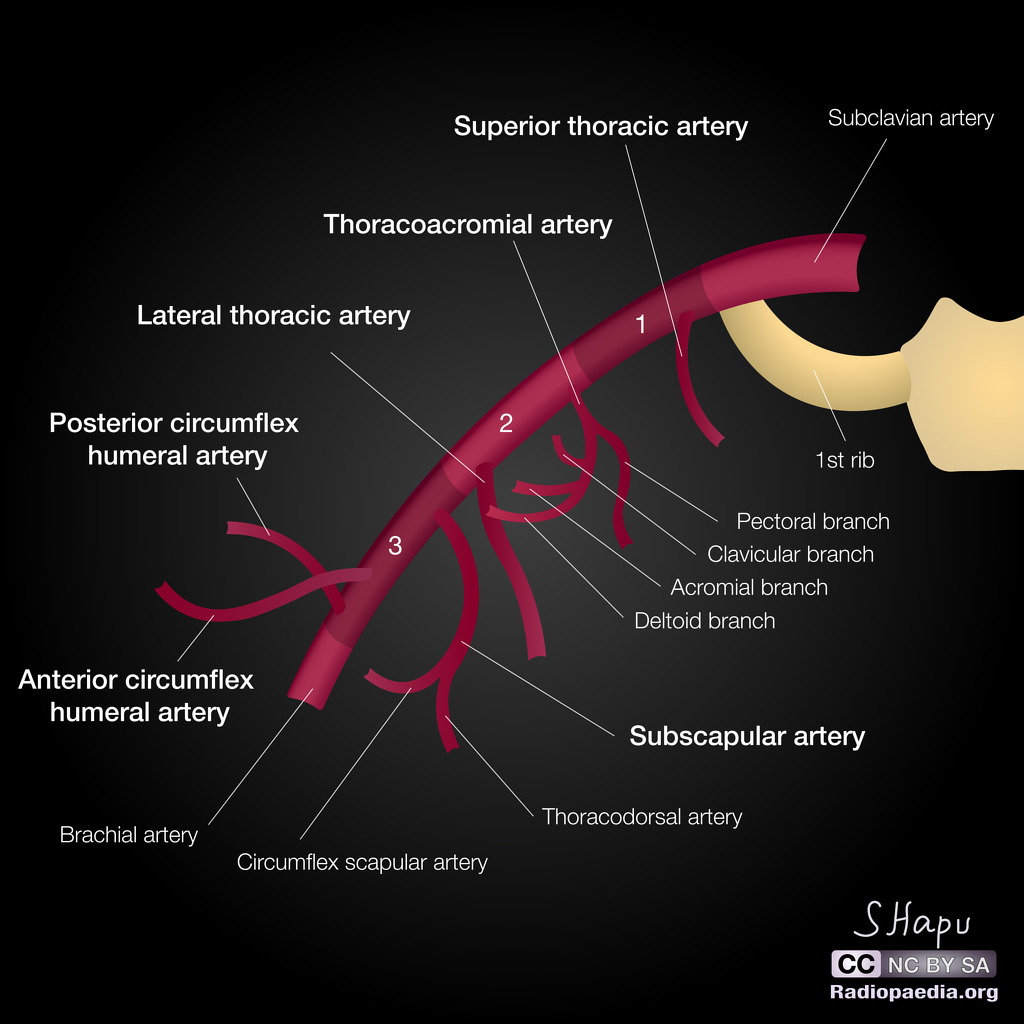
AXILLARY ARTERY
SUMMARY
ORIGIN
1. Continuation of the 3rd part of the subclavian a.
2. Begins at the outer border of the 1st rib, behind the midpoint of the clavicle.
COURSE & PARTS
3. At the lower border of the teres major it becomes the brachial a.
4. It is invested by the axillary sheath, continuation of the prevertebral fascia.
5. Divided into 3 parts by the pectoralis minor: first part (above), second part (behind), third part (below).
6. The cords of the brachial plexus are named according to their relations to the second part of the axillary artery.
7. The subclavian vein lies medially throughout its course.

Image: Case courtesy of Dr Sachintha Hapugoda, Radiopaedia.org. From the case rID: 52195 [Accessed 24 Nov. 2018].
Reference(s)
R.M.H McMinn (1998). Last’s anatomy: regional and applied. Edinburgh: Churchill Livingstone.
Gray, H., Carter, H.V. and Davidson, G. (2017). Gray’s anatomy. London: Arcturus.