CPPD DEPOSITION DISEASE-INVESTIGATION
From NeuroRehab.wiki
Revision as of 11:30, 1 January 2023 by Dr Appukutty Manickam (talk | contribs) (Imported from text file)
SUMMARY
1. Diagnose CPPD by finding intracellular crystals that are blunted, rhomboid, and are weakly positively birefringent.
2. Joint-space narrowing.
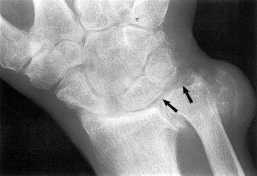
3. Chondrocalcinosis: ossification of the cartilage (see image below). Typical of pseudogout.

4. Cyst formation under the cartilage.
Reference(s)
Wilkinson, I. (2017). Oxford handbook of clinical medicine. Oxford: Oxford University Press.
Hannaman, R. A., Bullock, L., Hatchell, C. A., & Yoffe, M. (2016). Internal medicine review core curriculum, 2017-2018. CO Springs, CO: MedStudy.
Therapeutic Guidelines. Melbourne: Therapeutic Guidelines Limited. https://www.tg.org.au [Accessed 2021].