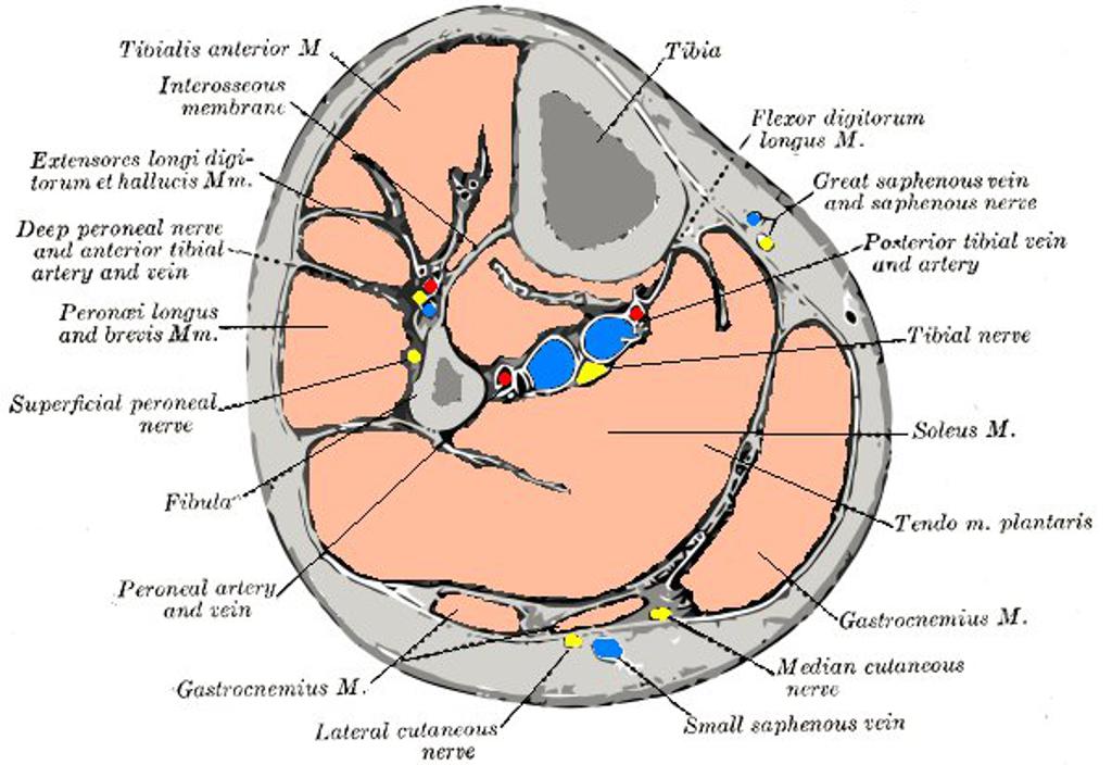
ANTERIOR COMPARTMENT OF LEG
From NeuroRehab.wiki
Revision as of 11:29, 1 January 2023 by Dr Appukutty Manickam (talk | contribs) (Imported from text file)
SUMMARY
1. Space b/w the deep fascia and interosseous membrane. 2. Bounded medially by the tibia and laterally by the fibula & anterior intermuscular septum.
3. 4 Muscles - tibialis anterior, extensor halucis longus, extensor digitorum longus, peroneus tertius. The tendons of these muscles pass beneath the superior extensor retinaculum to enter the foot.
4. Others - deep peroneal nerve, anterior tibial vessels.

Image: Case courtesy of Dr Jeremy Jones, Radiopaedia.org. From the case rID: 36325 [Accessed 12 Apr. 2019].
Reference(s)
R.M.H McMinn (1998). Last’s anatomy: regional and applied. Edinburgh: Churchill Livingstone.
Gray, H., Carter, H.V. and Davidson, G. (2017). Gray’s anatomy. London: Arcturus.