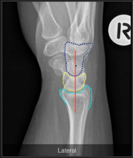
RADIOGRAPH-LATERAL WRIST
From NeuroRehab.wiki
Revision as of 11:29, 1 January 2023 by Dr Appukutty Manickam (talk | contribs) (Imported from text file)
SUMMARY
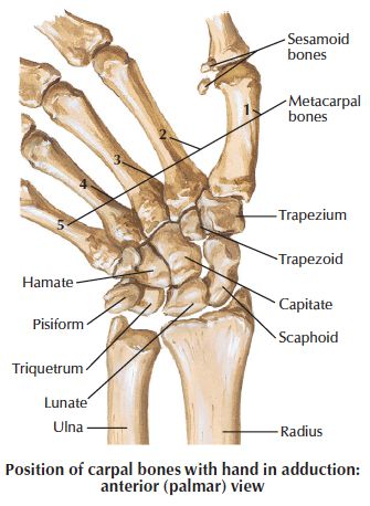
1. Remember that you do not see the first row of bones - scaphoid & trapezium, but the second row - lunate & capitate.
2. The lunate appears as a moon shaped bone.
3. Important to detect SCAPHO-LUNATE DISLOCATION.


Image: Case courtesy of Dr Andrew Dixon, Radiopaedia.org. From the case rID: 43305 [Accessed 18 Apr. 2020].
Reference(s)
R.M.H McMinn (1998). Last’s anatomy: regional and applied. Edinburgh: Churchill Livingstone.
Gray, H., Carter, H.V. and Davidson, G. (2017). Gray’s anatomy. London: Arcturus.