MRI-DIFFUSION
SUMMARY
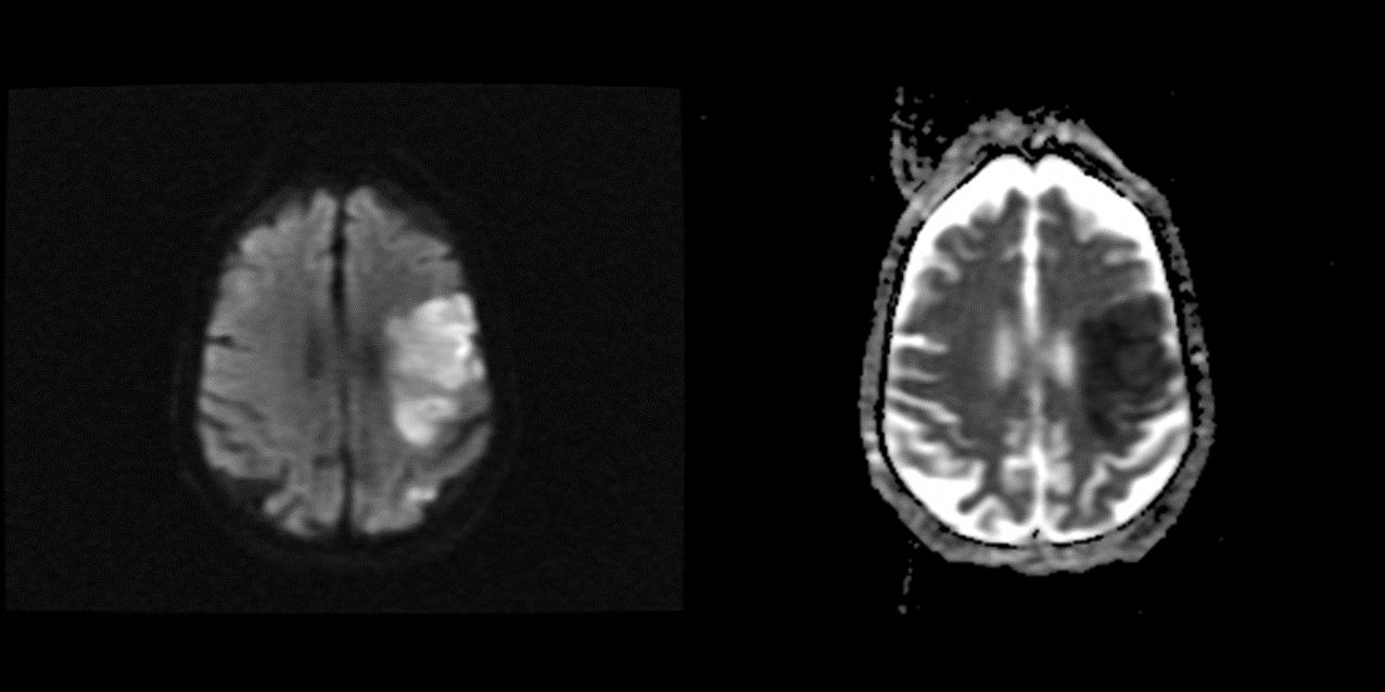
When describing diffusion weighted sequences, we also use the term intensity but additionally we use the terms "restricted diffusion" and "facilitated diffusion" to denote whether water can move around less easily (restricted) or more easily (facilitated) than expected for that tissue.

Images: The above reveal a region of hyperintensity on DWI on the left, wheres the same region demonstrates restricted diffusion on ADC mapping on the right; this is suggesteive of ischemic infarction.
Reference(s)
Furman, Michael B., and Leland Berkwits. Atlas of Image-Guided Spinal Procedures. Elsevier, Inc, 2017.
Horowitz AL. MRI Physics for Physicians. Springer Science & Business Media. (1989) ISBN:1468403338.
Mangrum W, Christianson K, Duncan S et-al. Duke Review of MRI Principles. Mosby. (2012) ISBN:1455700843.