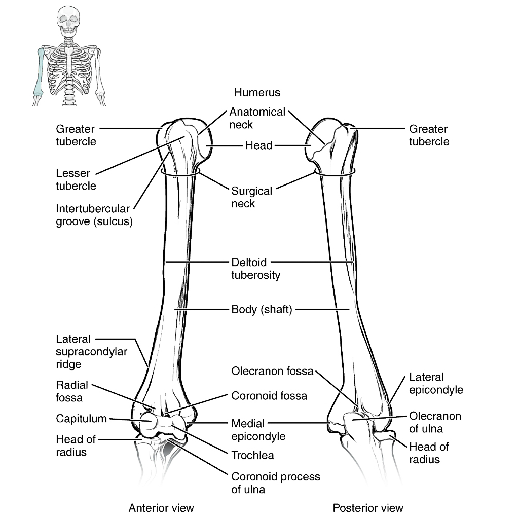
OSTEOLOGY-HUMERUS (UPPER PART)
SUMMARY
1. Superior articular head (forms third of a sphere) and articular margin is the anatomical neck. 2. Surgical neck - lies below the epiphyseal line, the axillary nerve winds around it.
3. Lesser tuberosity/tubercle - projects forward and is continued downwards as the medial lip of the bicipital groove.
4. Bicipital/intertubercular groove - lies anteriorly b/w the greater and lesser tuberosities. The LONG HEAD OF THE BICEPS TENDON passes through this, bridged by the transverse humeral ligament. The floor receives the tendon of the LATISSIMUS DORSI.
5. Greater tuberosity - has 3 facets for the SUPRASPINATUS, INFRASPINATUS, TERES MINOR.
6. The lateral lip of the bicipital groove runs into the anterior margin of the deltoid tuberosity and receives the PACTORALIS MAJOR TENDON.

Image: Case courtesy of OpenStax College, Radiopaedia.org. From the case rID: 42766 [Accessed 17 Apr. 2019].
Reference(s)
R.M.H McMinn (1998). Last’s anatomy: regional and applied. Edinburgh: Churchill Livingstone.
Gray, H., Carter, H.V. and Davidson, G. (2017). Gray’s anatomy. London: Arcturus.