DEEP POSTERIOR COMPARTMENT OF LEG-TIBIALIS POSTERIOR
From NeuroRehab.wiki
Revision as of 11:29, 1 January 2023 by Dr Appukutty Manickam (talk | contribs) (Imported from text file)
SUMMARY
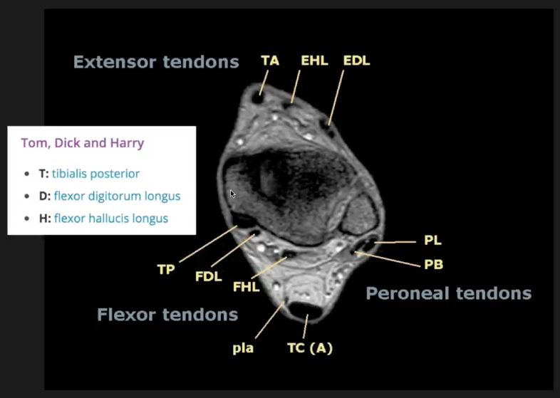
1. O: posterior tibia (below soleal line), posterior interosseous membrane & posterior fibula.
2. I: its tendon grooves the back of the medial malleolus, passes above the sustentaculum tali to be inserted into the navicular tuberosity & 3 cuneiforms.
3. NS: tibial n.
4. A: plantar flexion & inversion of foot.


Image: Gray, Henry. Anatomy of the Human Body. Philadelphia: Lea & Febiger, 1918; Bartleby.com, 2000. www.bartleby.com/107/ [Accessed 7 Apr. 2019].
Reference(s)
R.M.H McMinn (1998). Last’s anatomy: regional and applied. Edinburgh: Churchill Livingstone.
Gray, H., Carter, H.V. and Davidson, G. (2017). Gray’s anatomy. London: Arcturus.