SUPERFICIAL FLEXORS OF FOREARM-PRONATOR TERES
From NeuroRehab.wiki
Revision as of 11:29, 1 January 2023 by Dr Appukutty Manickam (talk | contribs) (Imported from text file)
SUMMARY
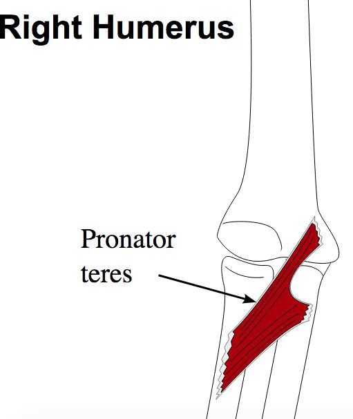
1. O: main head - medial epicondyle of humerus (common flexor origin) & medial supracondylar ridge. Deep head - medial border of the coronoid process.
2. I: lateral convexity of radial shaft.
3. NS: median n.
4. A: pronates forearm & flexes elbow.

Image: Egmason [CC BY-SA 3.0], via Wikimedia Commons [Accessed 20 Apr. 2019].
Reference(s)
R.M.H McMinn (1998). Last’s anatomy: regional and applied. Edinburgh: Churchill Livingstone.
Gray, H., Carter, H.V. and Davidson, G. (2017). Gray’s anatomy. London: Arcturus.