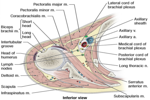
AXILLA-BOUNDARIES
SUMMARY
1. FLOOR - axillary fascia extending from the fascia over the SERRATUS ANTERIOR to the deep fascia of the arm. Attached in front & behind to the axillary folds. Suspended by a ligament from the lower border of PACTORALIS MINOR.
2. ANTERIOR WALL - PACTORALIS MAJOR, PACTORALIS MINOR, SUBCLAVIUS, clavipectoral fascia.
3. POSTERIOR WALL - SUBSCAPULARIS, TERES MAJOR, tendon of the LATISSIMUS DORSI.
4. MEDIAL WALL - SERRATUS ANTERIOR.
5. LATERAL WALL - humerus.
6. APEX - clavicle (anterior), scapula (posterior), first rib (medial). Channel of communication b/w axilla & posterior triangle of the neck.

Image: Tenderness.co. (2018). Axillary Artery. [online] Available at: http://tenderness.co/axillary-artery/ [Accessed 16 Nov. 2018].
Reference(s)
R.M.H McMinn (1998). Last’s anatomy: regional and applied. Edinburgh: Churchill Livingstone.
Gray, H., Carter, H.V. and Davidson, G. (2017). Gray’s anatomy. London: Arcturus.