Difference between revisions of "OSTEOLOGY-HUMERUS (LOWER PART)"
(Imported from text file) |
(Imported from text file) |
||
| Line 1: | Line 1: | ||
===== [[Summary Article|'''SUMMARY''']] ===== | ===== [[Summary Article|'''SUMMARY''']] ===== | ||
1. Nutrient foramen | <i>1. Nutrient foramen:</i> at the lower end of the <i>deltoid tuberosity, </i>on the medial aspect of the humerus. | ||
<br/>2. The | <br/> | ||
<br/>3. The | <br/>2. The coracobrachialis is inserted above this foramen. | ||
<br/> | |||
<br/>3. The lateral head of the triceps inserts into the lateral lip of the <i>radial groove. </i> | |||
<br/> | |||
<br/>4. The articular surface of the lower end of the humerus is coated with hyaline cartilage and shows the <i>capitulum (lateral) & trochlea (medial). </i> | <br/>4. The articular surface of the lower end of the humerus is coated with hyaline cartilage and shows the <i>capitulum (lateral) & trochlea (medial). </i> | ||
<br/><i>5. Capitulum | <br/> | ||
<br/><i>6. Trochlea | <br/><i>5. Capitulum: </i>for articulation with the head of the radius, arises laterally, projects down & forwards. | ||
<br/><i>7. Radial fossa | <br/> | ||
<br/><i>8. Coronoid fossa | <br/><i>6. Trochlea: </i>extends posteriorly. The lateral ridge of the trochlea is lower, produces a tilt at the lower end of the humerus and accounts for the carrying angle of the elbow. | ||
<br/>9. Medial epicondyle | <br/> | ||
<br/>10. Lateral epicondyle | <br/><i>7. Radial fossa: </i>anterior surface, above the capitulum. Accomodates the radial head in full flexion. | ||
<br/> | |||
<br/><i>8. Coronoid fossa: </i>anterior surface, above the trochlea. | |||
<br/> | |||
<br/><i>9. Medial epicondyle:</i> common flexor origin. | |||
<br/> | |||
<br/><i>10. Lateral epicondyle:</i> common extensor origin. | |||
<br/> | |||
<br/>[[Image:humerus.jpg]] | <br/>[[Image:humerus.jpg]] | ||
<br/> | <br/> | ||
Latest revision as of 18:52, 8 January 2023
SUMMARY
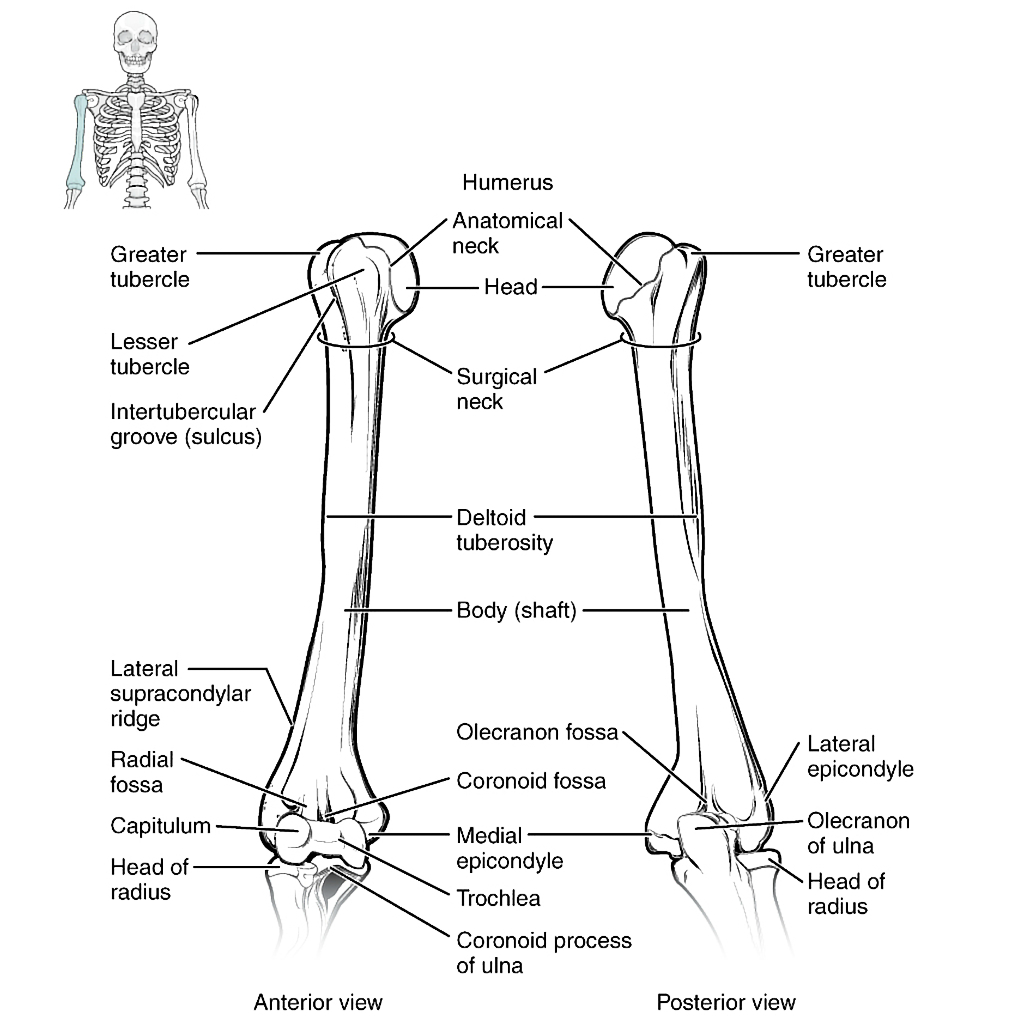
1. Nutrient foramen: at the lower end of the deltoid tuberosity, on the medial aspect of the humerus.
2. The coracobrachialis is inserted above this foramen.
3. The lateral head of the triceps inserts into the lateral lip of the radial groove.
4. The articular surface of the lower end of the humerus is coated with hyaline cartilage and shows the capitulum (lateral) & trochlea (medial).
5. Capitulum: for articulation with the head of the radius, arises laterally, projects down & forwards.
6. Trochlea: extends posteriorly. The lateral ridge of the trochlea is lower, produces a tilt at the lower end of the humerus and accounts for the carrying angle of the elbow.
7. Radial fossa: anterior surface, above the capitulum. Accomodates the radial head in full flexion.
8. Coronoid fossa: anterior surface, above the trochlea.
9. Medial epicondyle: common flexor origin.
10. Lateral epicondyle: common extensor origin.

Image: Case courtesy of OpenStax College, Radiopaedia.org. From the case rID: 42766 [Accessed 17 Apr. 2019].
Reference(s)
R.M.H McMinn (1998). Last’s anatomy: regional and applied. Edinburgh: Churchill Livingstone.
Gray, H., Carter, H.V. and Davidson, G. (2017). Gray’s anatomy. London: Arcturus.