Difference between revisions of "MUSCLES-ANTERIOR COMPARTMENT OF ARM"
(Imported from text file) |
(Imported from text file) |
||
| Line 1: | Line 1: | ||
[[Summary Article| | ===== [[Summary Article|'''SUMMARY''']] ===== | ||
1. CORACOBRACHIALIS - arises from the apex of the coracoid, attaches to the medial border of the humerus. <i>The musculocutaneous n.</i> runs through the muscle. Functionally unimportant weak flexor & adductor of the shoulder. | |||
<br/> | |||
<br/>2. BICEPS - <i>long head: </i>arises from the supraglenoid tubercle, passes through the capsule of the shoulder joint. <i>Short head: </i>arises from the apex of the coracoid. Its tendon inserts into the radial tubersoity & <i>bicipital aponeurosis </i>inserts into the subcutaneous border of proximal ulna.<i> Flexes the elbow & shoulder joints, supinates the forearm. </i> | <br/>2. BICEPS - <i>long head: </i>arises from the supraglenoid tubercle, passes through the capsule of the shoulder joint. <i>Short head: </i>arises from the apex of the coracoid. Its tendon inserts into the radial tubersoity & <i>bicipital aponeurosis </i>inserts into the subcutaneous border of proximal ulna.<i> Flexes the elbow & shoulder joints, supinates the forearm. </i> | ||
<br/ | <br/> | ||
<br/>3. BRACHIALIS - arises from the lower anterior 2/3 of the humerus & medial intermuscular septum. Inserted into the coronoid process & ulna tuberosity. <i>Flexes the elbow joint. </i> | <br/>3. BRACHIALIS - arises from the lower anterior 2/3 of the humerus & medial intermuscular septum. Inserted into the coronoid process & ulna tuberosity. <i>Flexes the elbow joint. </i> | ||
<br/ | <br/> | ||
<br/>4. BRACHIORADIALIS - arises from the upper 2/3 of the lateral supracondylar ridge. <i>Inserts into the radial styloid. The muscle overlies the radial nerve & artery. Flexes the elbow. Supplied by the radial nerve.</i> | <br/>4. BRACHIORADIALIS - arises from the upper 2/3 of the lateral supracondylar ridge. <i>Inserts into the radial styloid. The muscle overlies the radial nerve & artery. Flexes the elbow. Supplied by the radial nerve.</i> | ||
<br/> | <br/> | ||
<br/><i>5. All muscles except BRACHIORADIALIS are s</i><i>upplied by the musculocutaneous n. In addition, the lateral part of the BRACHIALIS is supplied by the radial n. </i> | <br/><i>5. All muscles except BRACHIORADIALIS are s</i><i>upplied by the musculocutaneous n. In addition, the lateral part of the BRACHIALIS is supplied by the radial n. </i> | ||
<br/ | <br/> | ||
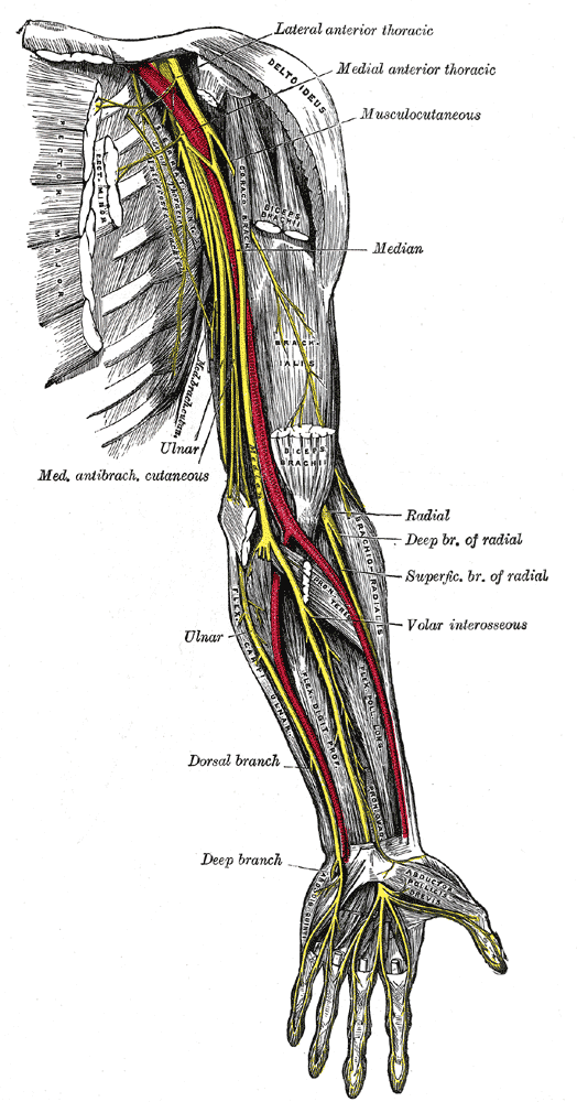
<br/>[[Image:Nerves_of_the_left_upper_extremity.gif]] | <br/>[[Image:Nerves_of_the_left_upper_extremity.gif]] | ||
<br/> | |||
<br/><b>Image:</b> [https://commons.wikimedia.org/wiki/File:Nerves_of_the_left_upper_extremity.gif Henry Vandyke Carter] / Public domain | <br/><b>Image:</b> [https://commons.wikimedia.org/wiki/File:Nerves_of_the_left_upper_extremity.gif Henry Vandyke Carter] / Public domain | ||
==Reference(s)== | ==Reference(s)== | ||
Revision as of 08:38, 30 December 2022
SUMMARY
1. CORACOBRACHIALIS - arises from the apex of the coracoid, attaches to the medial border of the humerus. The musculocutaneous n. runs through the muscle. Functionally unimportant weak flexor & adductor of the shoulder.
2. BICEPS - long head: arises from the supraglenoid tubercle, passes through the capsule of the shoulder joint. Short head: arises from the apex of the coracoid. Its tendon inserts into the radial tubersoity & bicipital aponeurosis inserts into the subcutaneous border of proximal ulna. Flexes the elbow & shoulder joints, supinates the forearm.
3. BRACHIALIS - arises from the lower anterior 2/3 of the humerus & medial intermuscular septum. Inserted into the coronoid process & ulna tuberosity. Flexes the elbow joint.
4. BRACHIORADIALIS - arises from the upper 2/3 of the lateral supracondylar ridge. Inserts into the radial styloid. The muscle overlies the radial nerve & artery. Flexes the elbow. Supplied by the radial nerve.
5. All muscles except BRACHIORADIALIS are supplied by the musculocutaneous n. In addition, the lateral part of the BRACHIALIS is supplied by the radial n.

Image: Henry Vandyke Carter / Public domain
Reference(s)
R.M.H McMinn (1998). Last’s anatomy: regional and applied. Edinburgh: Churchill Livingstone.
Gray, H., Carter, H.V. and Davidson, G. (2017). Gray’s anatomy. London: Arcturus.