Difference between revisions of "AXILLARY ARTERY-BRANCHES"
(Imported from text file) |
(Imported from text file) |
||
| Line 1: | Line 1: | ||
[[Summary Article| | ===== [[Summary Article|'''SUMMARY''']] ===== | ||
<b><i>TIP: mnemonic (Screw The Lawyer, Save A Patient!)</i></b> | |||
<br/ | <br/> | ||
<br/><i>1. First part:</i> | <br/><i>1. First part:</i> | ||
<br/><b>- S</b>uperior thoracic a. Supplies both pectoral muscles. <i>2. Second part:</i> | <br/><b>- S</b>uperior thoracic a. Supplies both pectoral muscles. | ||
<br/> | |||
<br/><i>2. Second part:</i> | |||
<br/><b>- T</b>horacoacromial a. (has 4 terminal branches) | <br/><b>- T</b>horacoacromial a. (has 4 terminal branches) | ||
<br/><b>- L</b>ateral thoracic a. (supplies both pectoral muscles & breast in women). | <br/><b>- L</b>ateral thoracic a. (supplies both pectoral muscles & breast in women). | ||
| Line 15: | Line 17: | ||
<br/> | <br/> | ||
<br/><b>Image:</b> Case courtesy of Dr Sachintha Hapugoda, [https://radiopaedia.org/ Radiopaedia.org]. From the case [https://radiopaedia.org/cases/52195 rID: 52195] [Accessed 16 Apr. 2019]. | <br/><b>Image:</b> Case courtesy of Dr Sachintha Hapugoda, [https://radiopaedia.org/ Radiopaedia.org]. From the case [https://radiopaedia.org/cases/52195 rID: 52195] [Accessed 16 Apr. 2019]. | ||
==Reference(s)== | ==Reference(s)== | ||
Revision as of 08:38, 30 December 2022
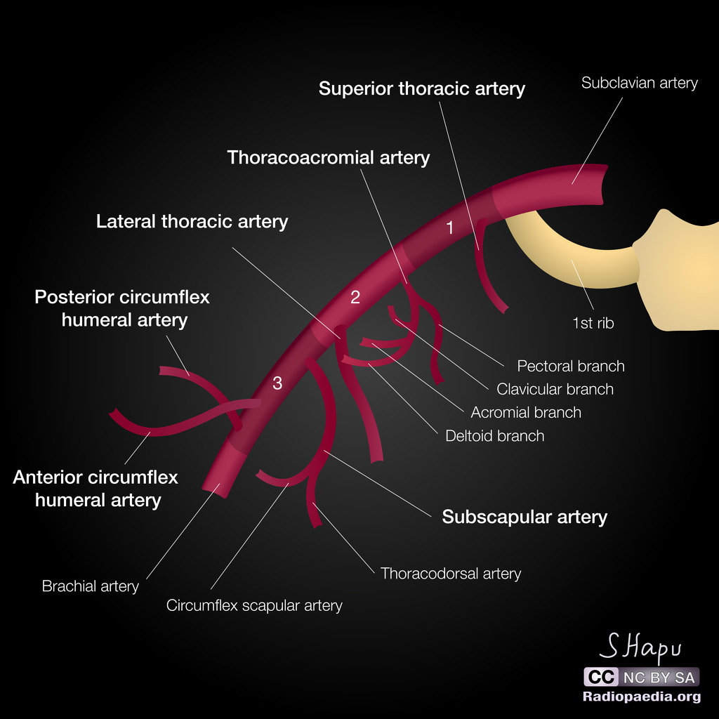
SUMMARY
TIP: mnemonic (Screw The Lawyer, Save A Patient!)
1. First part:
- Superior thoracic a. Supplies both pectoral muscles.
2. Second part:
- Thoracoacromial a. (has 4 terminal branches)
- Lateral thoracic a. (supplies both pectoral muscles & breast in women).
3. Third part:
- Subscapular a. (gives the circumflex scapular a. and changes its name to the thoracodorsal a.)
- Anterior circumflex humeral a. (anastamoses with the posterior circumflex humeral a. at the surgical neck of the humerus)
- Posterior circumflex humeral a. (accompanies the axillary n. through the quadrangular space to supply the deltoid).

Image: Case courtesy of Dr Sachintha Hapugoda, Radiopaedia.org. From the case rID: 52195 [Accessed 16 Apr. 2019].
Reference(s)
R.M.H McMinn (1998). Last’s anatomy: regional and applied. Edinburgh: Churchill Livingstone.
Gray, H., Carter, H.V. and Davidson, G. (2017). Gray’s anatomy. London: Arcturus.