Difference between revisions of "CONTENTS OF THE FEMORAL TRIANGLE"
From NeuroRehab.wiki
(Imported from text file) |
(Imported from text file) |
||
| Line 1: | Line 1: | ||
[[Summary Article|<h5>'''SUMMARY'''</h5>]] | [[Summary Article|<h5>'''SUMMARY'''</h5>]] | ||
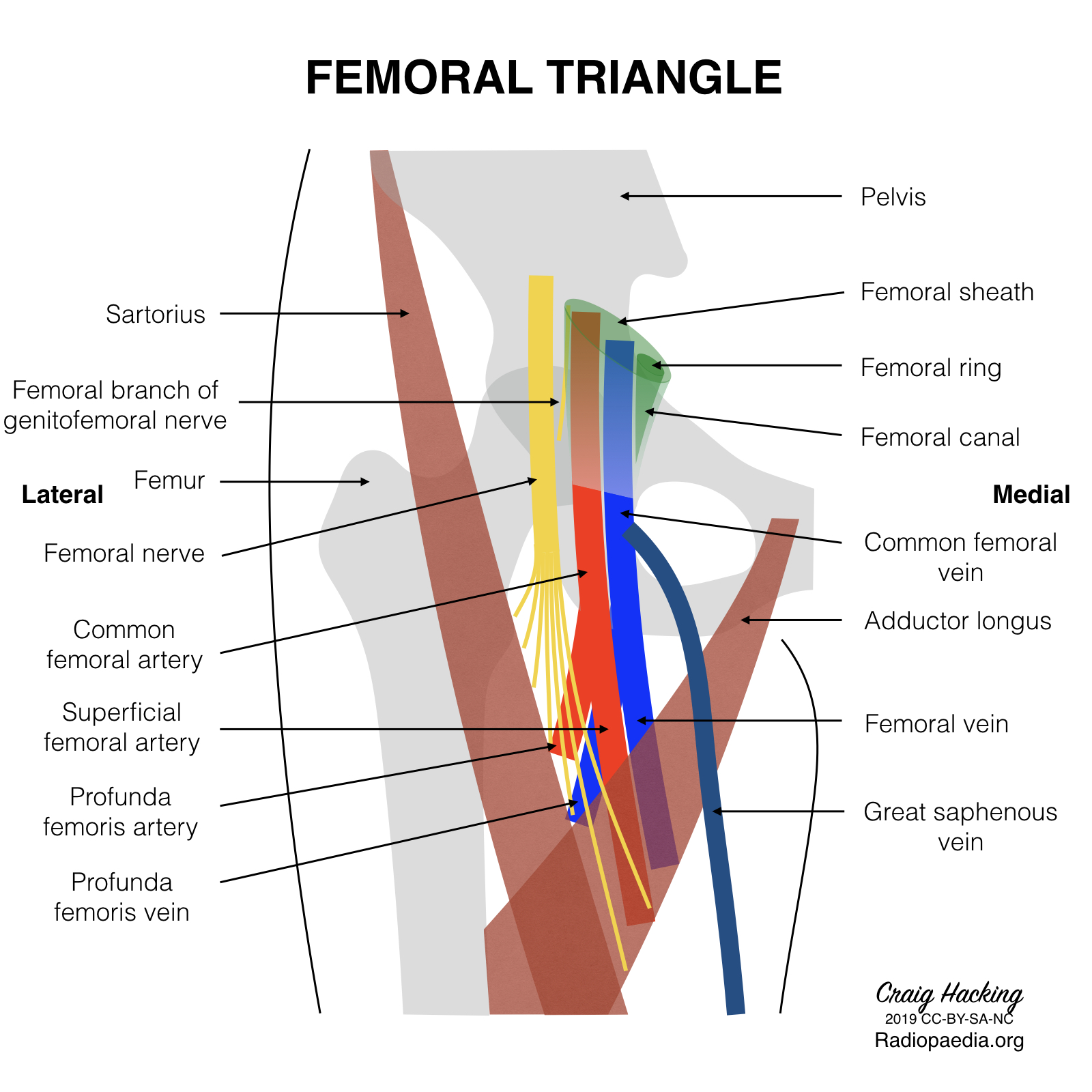
<br/><i>From lateral to medial: nerve => artery => vein => femoral canal (containing the deep inguinal LN). The nerve sits OUTSIDE the sheath. </i | <br/><i>From lateral to medial: nerve => artery => vein => femoral canal (containing the deep inguinal LN). The nerve sits OUTSIDE the sheath. </i>[[Image:femoral-triangle-diagram.jpeg]] | ||
<br/><b>Image: </b>Case courtesy of Dr Craig Hacking, [https://radiopaedia.org/ Radiopaedia.org]. From the case [https://radiopaedia.org/cases/70536 rID: 70536] | <br/><b>Image: </b>Case courtesy of Dr Craig Hacking, [https://radiopaedia.org/ Radiopaedia.org]. From the case [https://radiopaedia.org/cases/70536 rID: 70536] | ||
Revision as of 12:45, 27 December 2022
SUMMARY
From lateral to medial: nerve => artery => vein => femoral canal (containing the deep inguinal LN). The nerve sits OUTSIDE the sheath. 
Image: Case courtesy of Dr Craig Hacking, Radiopaedia.org. From the case rID: 70536
Reference(s)
R.M.H McMinn (1998). Last’s anatomy: regional and applied. Edinburgh: Churchill Livingstone.
Gray, H., Carter, H.V. and Davidson, G. (2017). Gray’s anatomy. London: Arcturus.