Difference between revisions of "ANTERIOR COMPARTMENT OF THIGH-VASTUS LATERALIS"
From NeuroRehab.wiki
(Imported from text file) |
(Imported from text file) |
||
| Line 1: | Line 1: | ||
[[Summary Article|<h5>'''SUMMARY'''</h5>]] | [[Summary Article|<h5>'''SUMMARY'''</h5>]] | ||
<br/>1. O: greater trochanter, lateral lip of the linea aspera, upper 2/3 of the lateral supracondylar line & | <br/>1. O: greater trochanter, lateral lip of the linea aspera, upper 2/3 of the lateral supracondylar line & lateral intermuscular septum. 2. I: base of patella & tibial tuberosity via patella tendon. | ||
<br/>3. NS: posterior branch of femoral n. | <br/>3. NS: posterior branch of femoral n. | ||
<br/>4. A: extends the knee. | <br/>4. A: extends the knee. | ||
Revision as of 12:45, 27 December 2022
SUMMARY
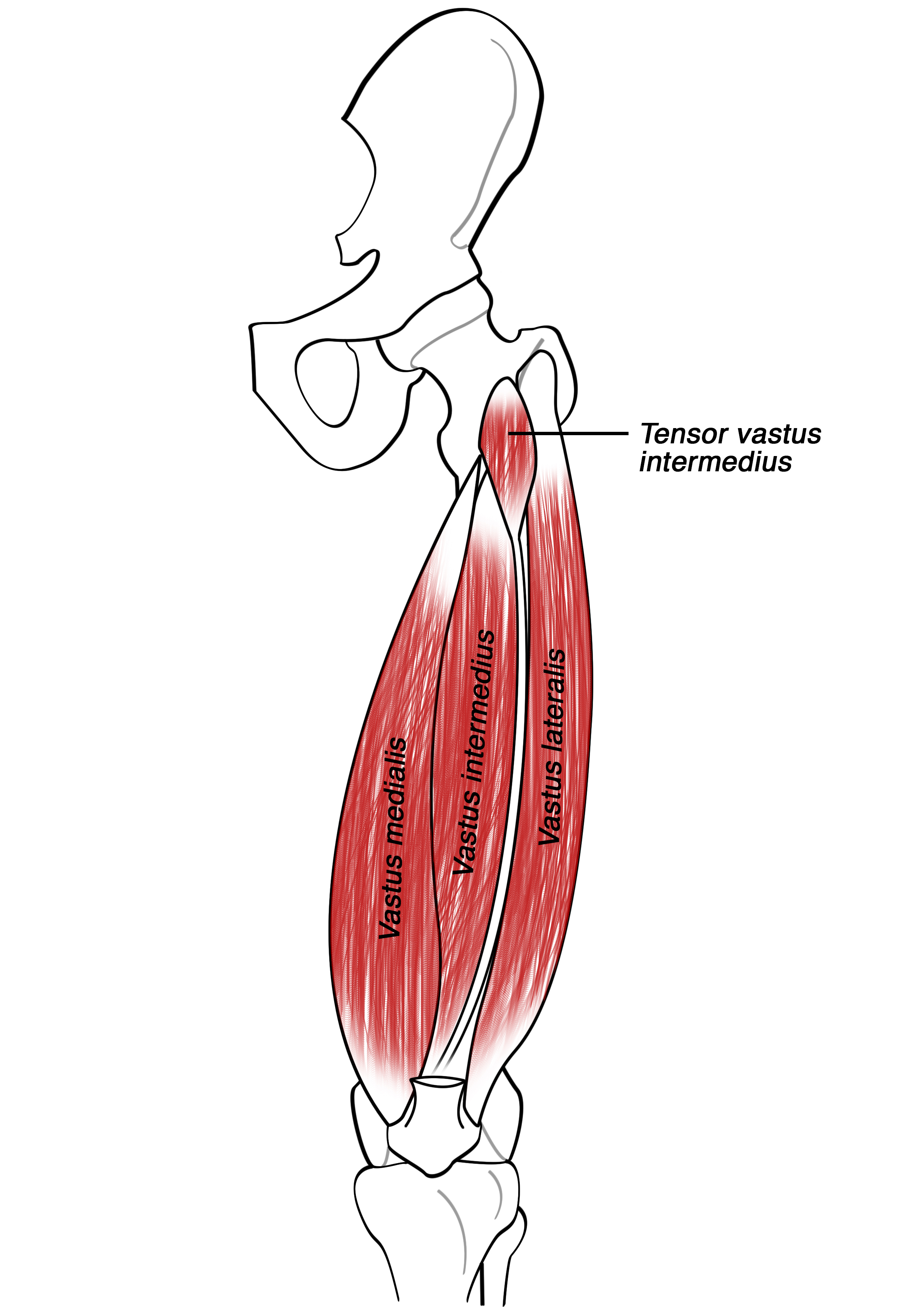
1. O: greater trochanter, lateral lip of the linea aspera, upper 2/3 of the lateral supracondylar line & lateral intermuscular septum. 2. I: base of patella & tibial tuberosity via patella tendon.
3. NS: posterior branch of femoral n.
4. A: extends the knee.

Image: Athikhun.suw [CC BY-SA 4.0], via Wikimedia Commons [Accessed 24 Sep. 2019].
Reference(s)
R.M.H McMinn (1998). Last’s anatomy: regional and applied. Edinburgh: Churchill Livingstone.
Gray, H., Carter, H.V. and Davidson, G. (2017). Gray’s anatomy. London: Arcturus.