Difference between revisions of "POSTERIOR CARPAL ARCH"
(Imported from text file) |
(Imported from text file) |
||
| Line 1: | Line 1: | ||
[[Summary Article|<h5>'''SUMMARY'''</h5>]] | [[Summary Article|<h5>'''SUMMARY'''</h5>]] | ||
<br/>1. Arterial anastamosis involving the radial a., ulnar a. & | <br/>1. Arterial anastamosis involving the radial a., ulnar a. & anterior interosseous a. | ||
<br/> | <br/> 2. Lies on the back of the carpus. | ||
<br/>3. Sends <i>dorsal metacarpal arteries</i> which run in the intermetacarpal spaces, deep to the long tendons. | <br/>3. Sends <i>dorsal metacarpal arteries</i> which run in the intermetacarpal spaces, deep to the long tendons. | ||
<br/>4. These split at the webs to supply the dorsal aspect of adjacent fingers. | <br/>4. These split at the webs to supply the dorsal aspect of adjacent fingers. | ||
Revision as of 12:45, 27 December 2022
SUMMARY
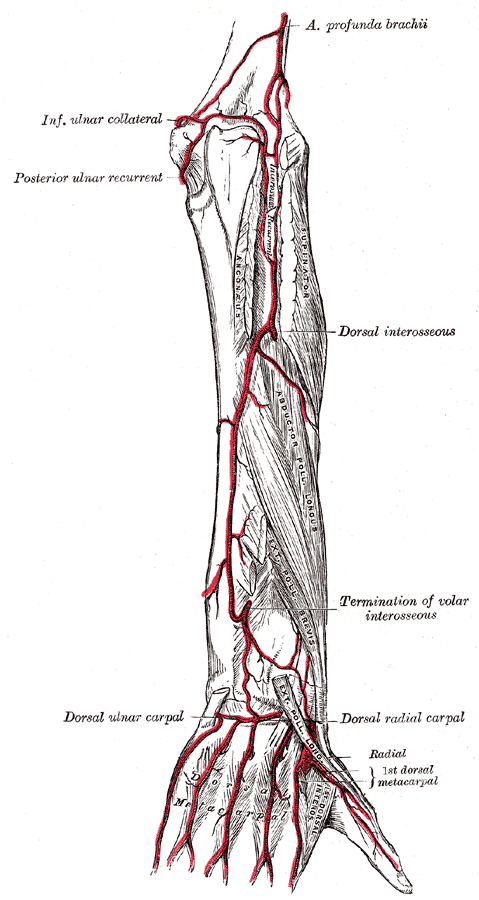
1. Arterial anastamosis involving the radial a., ulnar a. & anterior interosseous a.
2. Lies on the back of the carpus.
3. Sends dorsal metacarpal arteries which run in the intermetacarpal spaces, deep to the long tendons.
4. These split at the webs to supply the dorsal aspect of adjacent fingers.
5. They communicate through the interosseous spaces with the palmar metacarpal arteries of the deep palmar arch.
6. Companion veins drain the palm into the dorsal venous network.

Image: Gray, Henry. Anatomy of the Human Body. Philadelphia: Lea & Febiger, 1918; Bartleby.com, 2000. www.bartleby.com/107/ [Accessed 16 Apr. 2019].
Reference(s)
R.M.H McMinn (1998). Last’s anatomy: regional and applied. Edinburgh: Churchill Livingstone.
Gray, H., Carter, H.V. and Davidson, G. (2017). Gray’s anatomy. London: Arcturus.