Difference between revisions of "MEDIAN NERVE-CARPAL TUNNEL"
(Imported from text file) |
(Imported from text file) |
||
| Line 1: | Line 1: | ||
[[Summary Article| | ===== [[Summary Article|'''SUMMARY''']] ===== | ||
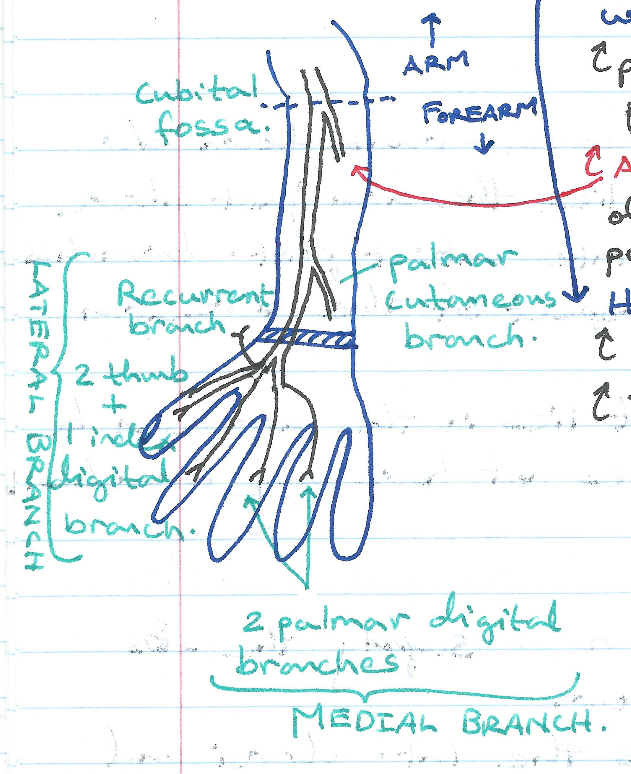
1. The median nerve gives off a <i>palmar cutaneous branch</i> as the main trunk emerges from underneath the FDS tendon approximately 3 to 4 cm before the distal wrist crease. | |||
<br/>2. This sensory branch usually exits from the radial (anterolateral surface) of the median nerve, travels <i>lateral</i> to the palmaris longus tendon and <i>superficial</i> to the flexor retinaculum to innervate the thenar eminence. | <br/>2. This sensory branch usually exits from the radial (anterolateral surface) of the median nerve, travels <i>lateral</i> to the palmaris longus tendon and <i>superficial</i> to the flexor retinaculum to innervate the thenar eminence. | ||
<br/>3. The median nerve most commonly passes through the tunnel without branching. | <br/>3. The median nerve most commonly passes through the tunnel without branching. | ||
| Line 11: | Line 11: | ||
<br/>[[Image:Median Nerve.jpg]] | <br/>[[Image:Median Nerve.jpg]] | ||
<br/><b>Image:</b> Dr. Appukutty Manickam. | <br/><b>Image:</b> Dr. Appukutty Manickam. | ||
==Reference(s)== | ==Reference(s)== | ||
Revision as of 08:38, 30 December 2022
SUMMARY
1. The median nerve gives off a palmar cutaneous branch as the main trunk emerges from underneath the FDS tendon approximately 3 to 4 cm before the distal wrist crease.
2. This sensory branch usually exits from the radial (anterolateral surface) of the median nerve, travels lateral to the palmaris longus tendon and superficial to the flexor retinaculum to innervate the thenar eminence.
3. The median nerve most commonly passes through the tunnel without branching.
4. It then gives off the recurrent motor branch just distal to the retinaculum on its radial side.
RECURRENT MOTOR BRANCH
5. The recurrent motor branch will then curve back to innervate the thenar muscles.
6. In 30% of cases the recurrent branch can arise on the ulnar side of the nerve and crosses over it towards its targets.
7. In 20% of cases it pierces the flexor retinaculum.

Image: Dr. Appukutty Manickam.
Reference(s)
R.M.H McMinn (1998). Last’s anatomy: regional and applied. Edinburgh: Churchill Livingstone.
Gray, H., Carter, H.V. and Davidson, G. (2017). Gray’s anatomy. London: Arcturus.