Difference between revisions of "MRI-MR PERFUSION"
(Imported from text file) |
|
(No difference)
| |
Latest revision as of 11:59, 25 July 2023
SUMMARY
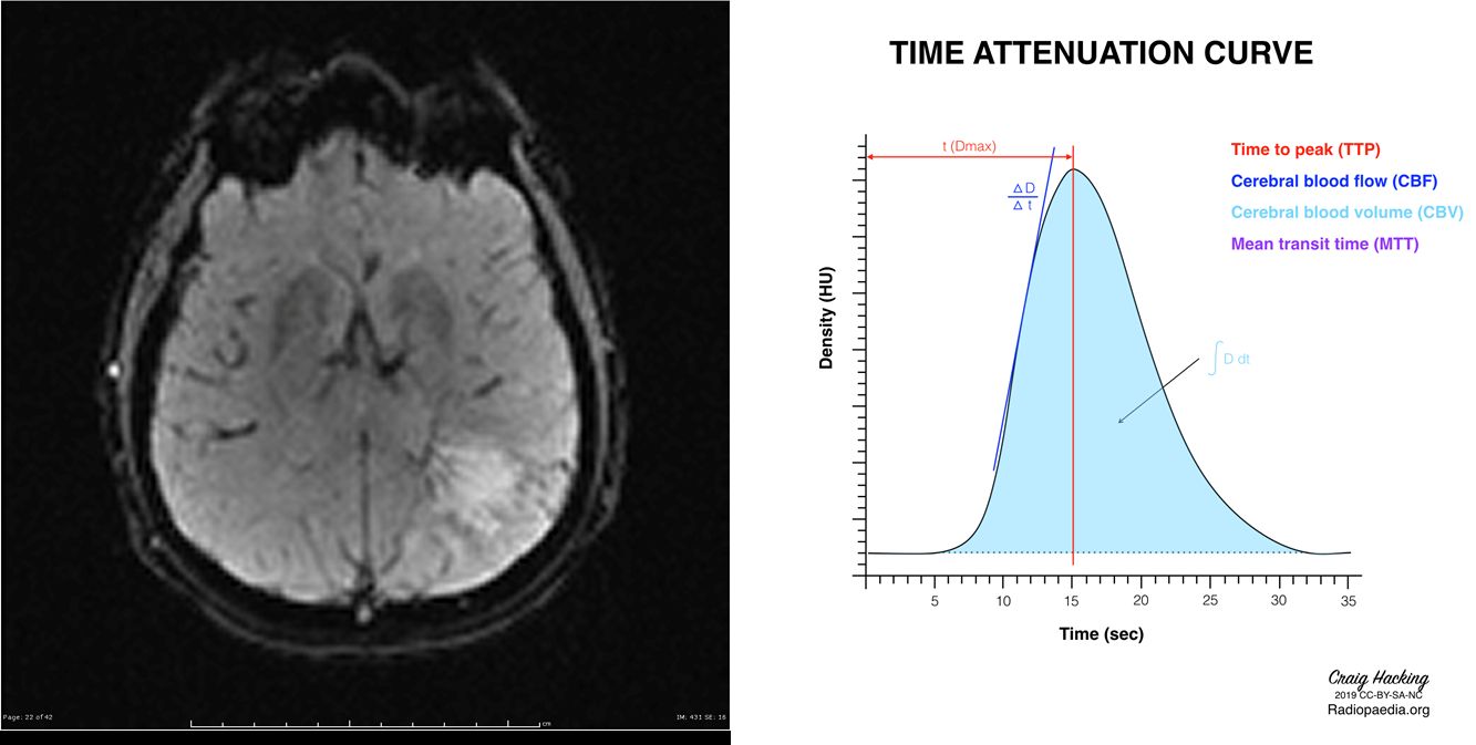
1.The amount of blood flowing into tissue can also be detected and relatively quantified, generating values such as cerebral blood volume, cerebral blood flow and mean transit time.
2.These values are useful in a number of clinical scenarios, including defining the ischaemic penumbra in ischaemic stroke, assessing histological grade of certain tumours, or distinguishing radionecrosis from tumour progression.

Image: Case courtesy of Craig Hacking, Radiopaedia.org. From the case rID: 70313.
Reference(s)
Furman, Michael B., and Leland Berkwits. Atlas of Image-Guided Spinal Procedures. Elsevier, Inc, 2017.
Horowitz AL. MRI Physics for Physicians. Springer Science & Business Media. (1989) ISBN:1468403338.
Mangrum W, Christianson K, Duncan S et-al. Duke Review of MRI Principles. Mosby. (2012) ISBN:1455700843.