Difference between revisions of "SPINE INJECTION-NEEDLE ADVANCEMENT"
(Imported from text file) |
(Imported from text file) |
||
| Line 11: | Line 11: | ||
<br/> | <br/> | ||
<br/><b>Images:</b> Furman, Michael B., and Leland Berkwits. <i>Atlas of Image-Guided Spinal Procedures</i>. Elsevier, Inc, 2017. | <br/><b>Images:</b> Furman, Michael B., and Leland Berkwits. <i>Atlas of Image-Guided Spinal Procedures</i>. Elsevier, Inc, 2017. | ||
==Reference(s)== | |||
Furman, Michael B., and Leland Berkwits. Atlas of Image-Guided Spinal Procedures. Elsevier, Inc, 2017. | |||
<br/>Horowitz AL. MRI Physics for Physicians. Springer Science & Business Media. (1989) ISBN:1468403338. | |||
<br/>Mangrum W, Christianson K, Duncan S et-al. Duke Review of MRI Principles. Mosby. (2012) ISBN:1455700843. | |||
[[Category:Spine Injection]] | [[Category:Spine Injection]] | ||
[[Category:Radiology]] | [[Category:Radiology]] | ||
[[Category:Radiology]] | [[Category:Radiology]] | ||
Revision as of 12:18, 25 April 2023
SUMMARY
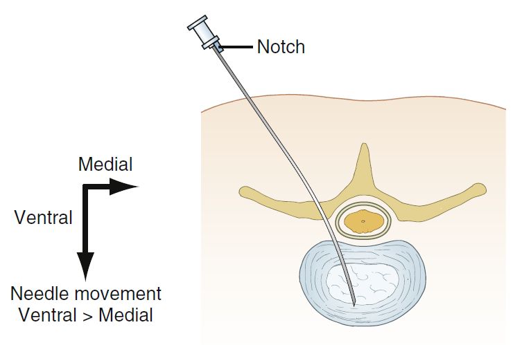
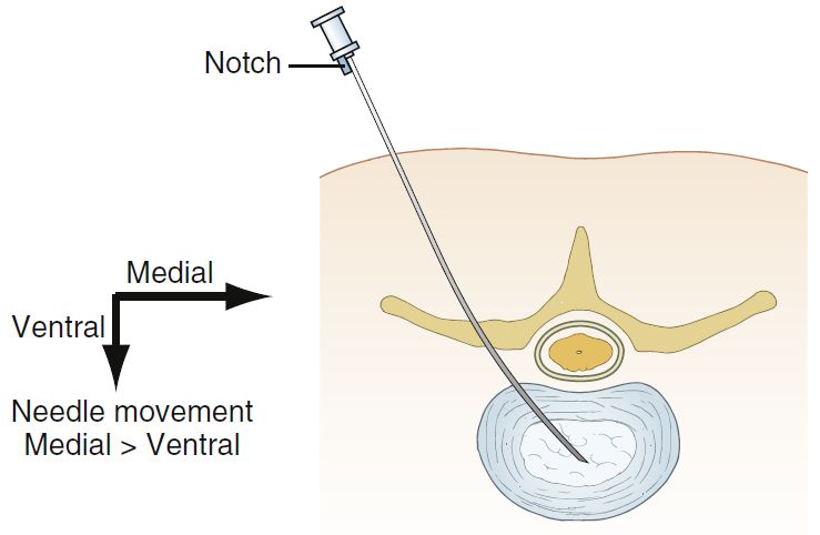
1. As a needle advances toward a target starting with an oblique projection, its trajectory can be broken into two predominant vectors: medial and ventral.
2. After a needle is embedded in a tissue and on its given path, placing the notch laterally will drive the needle tip medially.

3. Placing the notch medially will drive the needle tip ventrally.

4. if a needle trajectory is less oblique and more perpendicular to the skin surface (Fig. 2.11), then a lateral needle tip movement can be achieved.
Images: Furman, Michael B., and Leland Berkwits. Atlas of Image-Guided Spinal Procedures. Elsevier, Inc, 2017.
Reference(s)
Furman, Michael B., and Leland Berkwits. Atlas of Image-Guided Spinal Procedures. Elsevier, Inc, 2017.
Horowitz AL. MRI Physics for Physicians. Springer Science & Business Media. (1989) ISBN:1468403338.
Mangrum W, Christianson K, Duncan S et-al. Duke Review of MRI Principles. Mosby. (2012) ISBN:1455700843.