Difference between revisions of "ADDUCTOR CANAL-LEAVING IT!"
From NeuroRehab.wiki
(Imported from text file) |
(Imported from text file) |
||
| Line 1: | Line 1: | ||
===== [[Summary Article|'''SUMMARY''']] ===== | ===== [[Summary Article|'''SUMMARY''']] ===== | ||
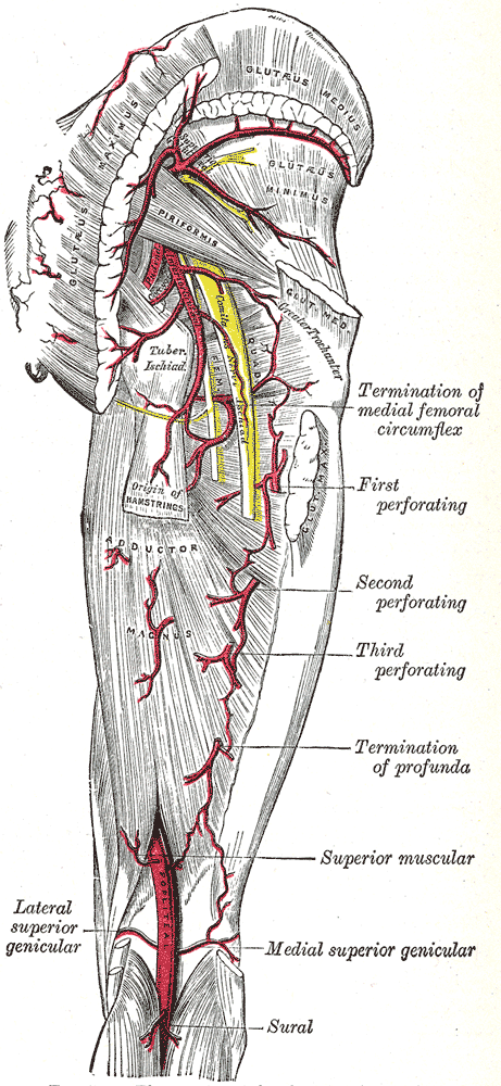
<i>The femoral vessels leave the femoral canal by passing into the popliteal fossa through a hiatus in the adductor magnus. | <i>The femoral vessels leave the femoral canal by passing into the popliteal fossa through a hiatus in the adductor magnus. </i> | ||
<br/>[[Image:Gray544.png]] | <br/>[[Image:Gray544.png]] | ||
<br/><b>Image: | <br/><b>Image: </b>Gray, Henry. <i>Anatomy of the Human Body.</i> Philadelphia: Lea & Febiger, 1918; Bartleby.com, 2000. [https://www.bartleby.com/107/ www.bartleby.com/107/] [Accessed 7 Apr. 2019]. | ||
Latest revision as of 11:29, 1 January 2023
SUMMARY
The femoral vessels leave the femoral canal by passing into the popliteal fossa through a hiatus in the adductor magnus.

Image: Gray, Henry. Anatomy of the Human Body. Philadelphia: Lea & Febiger, 1918; Bartleby.com, 2000. www.bartleby.com/107/ [Accessed 7 Apr. 2019].
Reference(s)
R.M.H McMinn (1998). Last’s anatomy: regional and applied. Edinburgh: Churchill Livingstone.
Gray, H., Carter, H.V. and Davidson, G. (2017). Gray’s anatomy. London: Arcturus.