Difference between revisions of "BRACHIAL ARTERY"
(Imported from text file) |
(Imported from text file) |
||
| Line 4: | Line 4: | ||
<br/> | <br/> | ||
<br/>RELATIONS | <br/>RELATIONS | ||
<br/>2. The axillary vein runs medial to the axillary artery, formed by the <i>venae comitantes of the brachial artery & basilic v. | <br/>2. The axillary vein runs medial to the axillary artery, formed by the <i>venae comitantes of the brachial artery & basilic v. </i> | ||
<br/>3. The median nerve runs lateral to it above, then cross over it to lie on its medial side below. | <br/> | ||
<br/>4. The ulnar nerve runs posterior to it above, then leaves it by piercing the medial intermuscular septum below. | <br/>3. The median nerve runs lateral to it above, then cross over it to lie on its medial side below. | ||
<br/>4. The ulnar nerve runs posterior to it above, then leaves it by piercing the medial intermuscular septum below. | |||
<br/> | <br/> | ||
<br/>COURSE | <br/>COURSE | ||
<br/>5. The artery is superficial throughout its course in the arm, in the groove b/w the biceps & triceps, and enters the cubital fossa before dividing into the <i>radial & ulnar arteries. | <br/>5. The artery is superficial throughout its course in the arm, in the groove b/w the biceps & triceps, and enters the cubital fossa before dividing into the <i>radial & ulnar arteries. </i> | ||
<br/> | |||
<br/><i>6. Surface marking - along a line from the bicipital groove to middle of the cubital fossa, at the level of the radial neck.</i> | <br/><i>6. Surface marking - along a line from the bicipital groove to middle of the cubital fossa, at the level of the radial neck.</i> | ||
<br/> | <br/> | ||
<br/>BRANCHES | <br/>BRANCHES | ||
<br/><i>7. Branches - radial a., ulnar a., profunda brachii a., superior ulnar collateral a., inferior ulnar collateral a., muscular branches, humeral nutrient branch. | <br/><i>7. Branches - radial a., ulnar a., profunda brachii a., superior ulnar collateral a., inferior ulnar collateral a., muscular branches, humeral nutrient branch. </i> | ||
<br/>[[Image:paste-13249974109060.jpg]] | <br/>[[Image:paste-13249974109060.jpg]] | ||
<br/><b>Image: | <br/><b>Image: </b>Gray, Henry. Anatomy of the Human Body. Philadelphia: Lea & Febiger, 1918; Bartleby.com, 2000. www.bartleby.com/107/ [Accessed 19 Nov. 2018]. | ||
Revision as of 11:29, 1 January 2023
SUMMARY
ORIGIN
1. This is the continuation of the third part of the axillary artery at the lower border of the TERES MAJOR.
RELATIONS
2. The axillary vein runs medial to the axillary artery, formed by the venae comitantes of the brachial artery & basilic v.
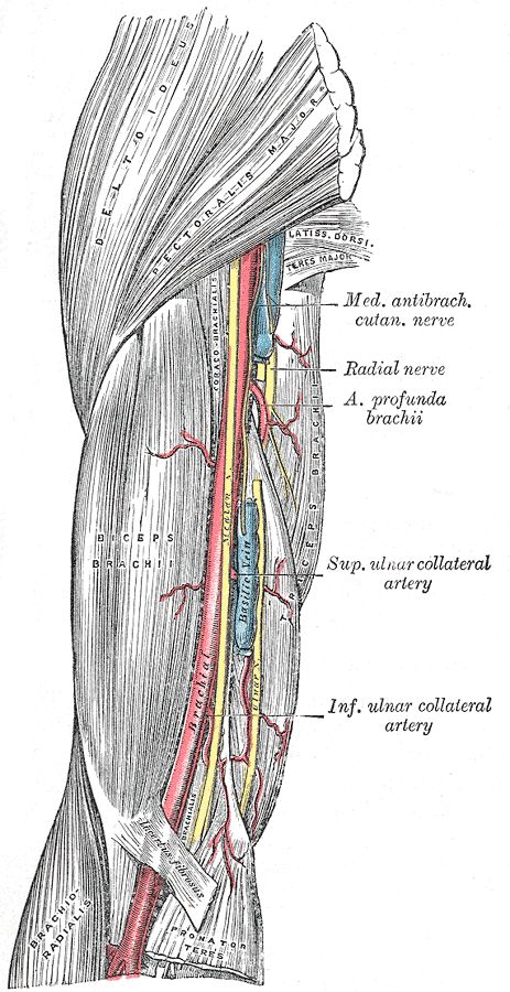
3. The median nerve runs lateral to it above, then cross over it to lie on its medial side below.
4. The ulnar nerve runs posterior to it above, then leaves it by piercing the medial intermuscular septum below.
COURSE
5. The artery is superficial throughout its course in the arm, in the groove b/w the biceps & triceps, and enters the cubital fossa before dividing into the radial & ulnar arteries.
6. Surface marking - along a line from the bicipital groove to middle of the cubital fossa, at the level of the radial neck.
BRANCHES
7. Branches - radial a., ulnar a., profunda brachii a., superior ulnar collateral a., inferior ulnar collateral a., muscular branches, humeral nutrient branch.

Image: Gray, Henry. Anatomy of the Human Body. Philadelphia: Lea & Febiger, 1918; Bartleby.com, 2000. www.bartleby.com/107/ [Accessed 19 Nov. 2018].
Reference(s)
R.M.H McMinn (1998). Last’s anatomy: regional and applied. Edinburgh: Churchill Livingstone.
Gray, H., Carter, H.V. and Davidson, G. (2017). Gray’s anatomy. London: Arcturus.